A Short Drug Exposure in Developing Organoids
An East Carolina University team generated hippocampal organoids from the IMR90-4 human induced pluripotent stem cell (iPSC) line. On day 57 of differentiation, the cultures received 1 mM valproic acid or no drug for four days. The researchers then sequenced messenger RNA (mRNA) and microRNA (miRNA) from the organoid groups to examine molecular changes associated with the exposure.
Valproic acid is an antiseizure and mood-stabilizing medicine. Its association with neurodevelopmental risk after prenatal exposure makes its molecular effects relevant to developmental drug-safety research.
Connecting the RNA Changes to Candidate Mechanisms
The study paired the two sequencing approaches because miRNAs can regulate the abundance or translation of messenger RNAs. The investigators first identified exposure-associated mRNA changes, then examined altered miRNAs and their predicted target relationships. This generated a candidate regulatory network rather than a direct measurement of every interaction.
Alongside genome-wide profiling, a focused analysis highlighted 60 genes associated with neuroinflammation, spanning inflammatory signaling, excitation–inhibition-related processes, developmental regulation, oxidative stress and neurovascular-associated genes. The researchers checked 10 selected transcripts by RT-qPCR. Their follow-up prioritized substantial expression changes, so it did not independently validate every proposed link in the regulatory network.
Functional annotation placed altered genes in immune-related processes, neuronal projections and potassium-channel biology. A synapse-focused analysis also identified processes associated with postsynaptic signaling and receptor localization. These results locate the RNA changes within known gene functions. The experiment did not record those inferred synaptic or ion-channel effects directly, and the organoid transcriptome does not show which of them would dominate a developing brain’s functional response.
Nineteen miRNAs and Selected Target Tests
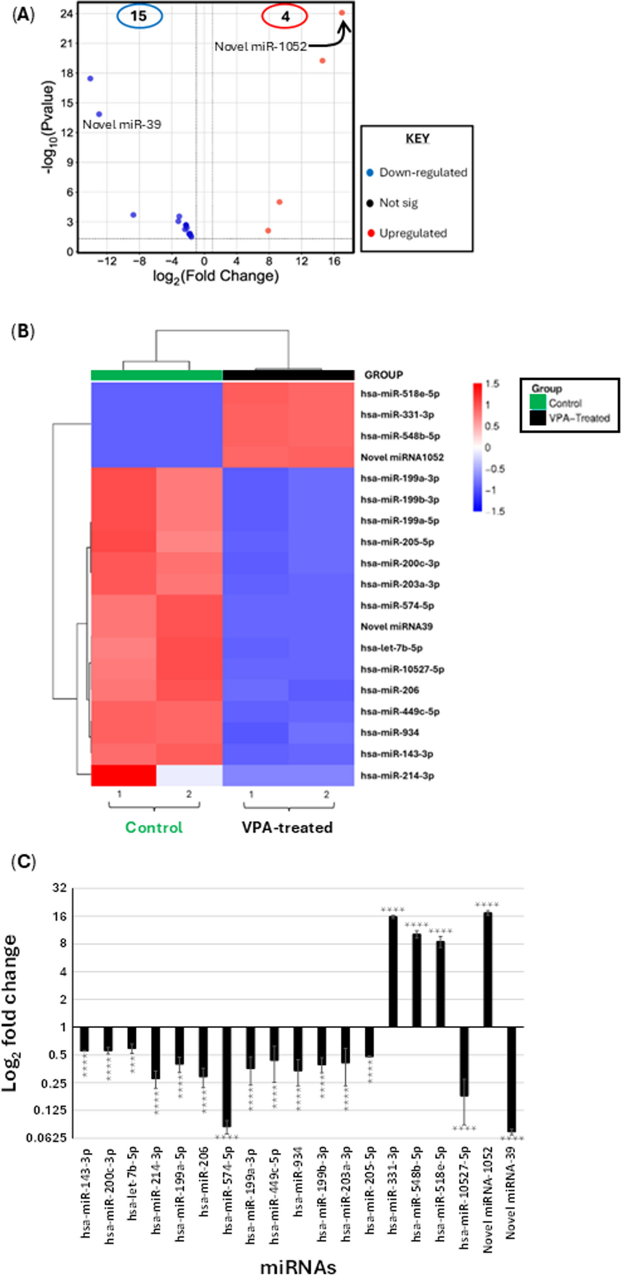
The miRNA analysis identified 19 miRNAs with altered expression after exposure: 17 known miRNAs and two new candidates named Novel miR-39 and Novel miR-1052. Across predicted miRNA–mRNA pairs, the overall inverse association was weak (Pearson r = −0.1086), leaving individual regulatory links to be tested.
In HEK-293T reporter cells, Novel miR-39 reduced activity for the TGFBR3 and GABRB2 constructs, while Novel miR-1052 did so for NDEL1 and GPBP1. The predicted HS6ST2 interaction did not produce a comparable reporter response. These sequence-level tests did not establish the magnitude of regulation inside the hippocampal organoids.
Effects in One iPSC Line
Sequencing used two biological replicates per group from a single female-donor iPSC line. This design limits estimates of biological variation and the detection of low-abundance miRNAs. The organoids lacked microglia and vasculature, so the reported inflammation-associated changes describe molecular signaling in the neural compartment.