C9ORF72 Patient-Neuron RNA-Foci Screen with RNA-Fate Counterassays
Screen patient-derived induced neurons for repeat-RNA foci modulation, filter general gene-expression effects, and follow RNA location and repeat-protein/growth outcomes in distinct mechanistic models.
Brain & Neural ModelsHuman Cell Assays
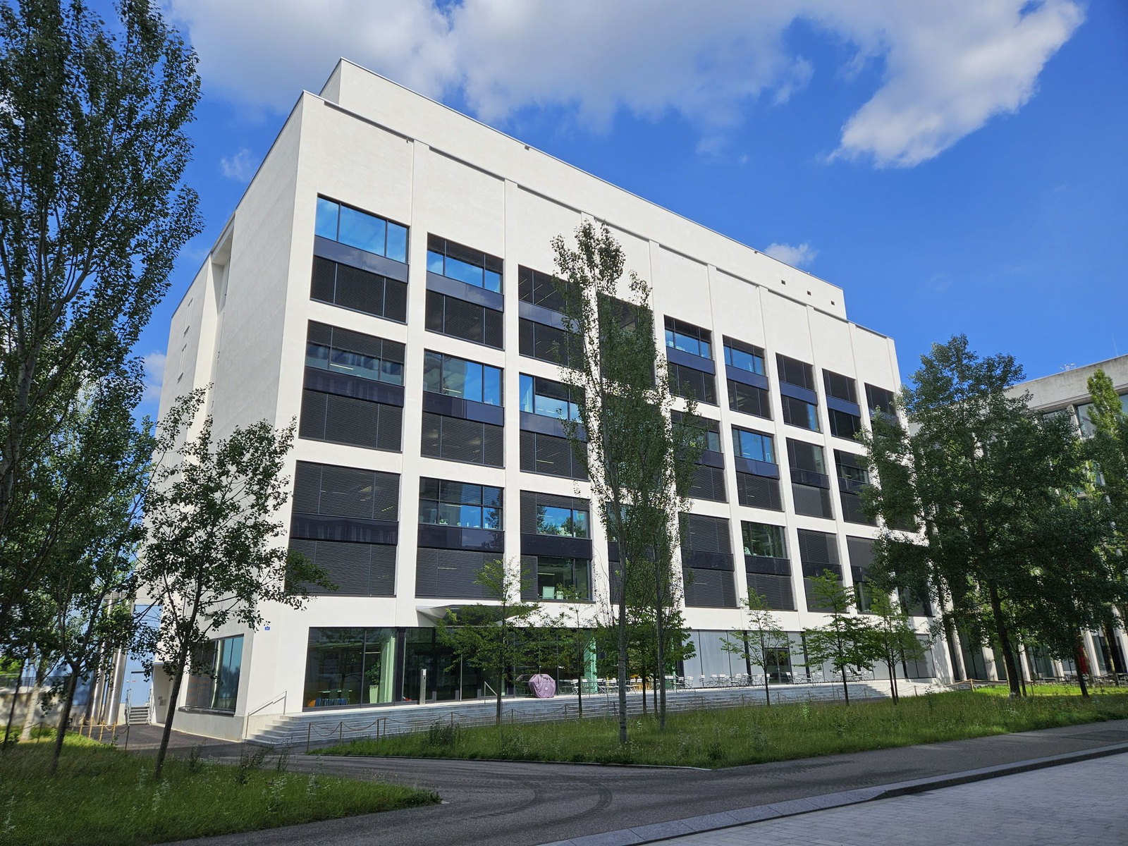
The Friedrich Miescher Institute’s St. Johann campus building in Basel, photographed in November 2024. (GP modernus, CC BY-SA 4.0)
Research Applications
Identify pharmacological tools for C9ORF72 repeat-RNA biology while testing whether foci clearance tracks a beneficial downstream response.
Model & Design
Primary ALS80/5a neurons induced from iPSCs using Neurogenin-2; additional C9ORF72 donor lines and motor neurons; separate engineered HeLa/HEK reporter systems for RNA fate, translation and growth.
Experimental Format
96,200 compounds at 8 μM for 48 hours in 1,536-well plates; single initial replicate; RNA-foci/nucleus imaging with at least 40% foci change and at most 40% cell-number reduction for initial hit selection; subsequent dose-response, counterassay and donor-line testing.
Model Components
Component
Primary readout
Description
Repeat-RNA fluorescence in situ hybridization and automated nuclear/spot counting; Acapella image-analysis software quantifies foci relative to cell numbers; healthy-donor and ALS vehicle controls on each plate.
Component
Follow-up tools
Description
SML-3, inactive SML-4 and orthogonal SF3B1 modulators; gene-expression reporters, RNA assays, single-molecule export imaging, tagged repeat-protein detection and live-cell growth tracking. Tagged poly-GA/GP imaging used 100 nM SML-3 or pladienolide B for 24 hours; growth comparisons used 100 nM SML-3/SML-4/pladienolide B over around four days.
Measurements & Endpoints
RNA foci per nucleus
Cell count and dose response
General transcription/translation interference
Nuclear/cytoplasmic repeat RNA
Reporter dipeptide-repeat levels
Reporter-cell proliferation
Analysis
Keep the primary single-line neuron screen, separate donor validation and engineered-reporter outcomes distinct. Foci reduction is not equated with RNA destruction or neuroprotection.
Published Evidence
Study Design
Retest primary hits, remove general-expression interferers, and probe active/inactive analogs, target depletion, RNA fate and reporter-protein/growth effects.
Study Sample
96,200 primary compounds; 2,952 dose-response candidates; 261 confirmed foci modulators; 122 general-expression interferers removed; 82 final hits after testing lines from three independent donors.
Comparison
Healthy-donor/ALS vehicle references, active versus inactive SML analogs, orthogonal SF3B1 compounds, protein depletion and induced/uninduced reporter controls.
Finding
SML-3 clears foci at about 2 nM in tested fibroblast/induced-neuron assays. Pharmacological SF3B1 modulation reduces foci while depletion increases them. At 100 nM, SML-3 or pladienolide B increased tagged poly-GA/GP after 24 hours; active-compound growth effects were tracked at the same nominal concentration over around four days in HEK reporters. Native neuronal dipeptide-repeat proteins could not be robustly detected.
Interpretation & Limits
Initial primary screen uses one neuron line and one replicate per compound.
Foci vary between donors/clones; final hits include both foci-increasing and foci-decreasing compounds.
Protein/growth readouts are engineered reporter outcomes, with tag effects and broad splicing changes as confounders.
Dipeptide-repeat increase correlates with growth inhibition; direct causality and native patient-neuron consequences were not established.
A 96,200-compound screen in patient-derived neurons found modulators of C9ORF72 repeat-RNA foci. Follow-up studies showed that some foci-clearing compounds increased repeat-protein production and reduced growth in engineered HEK reporter cells, complicating interpretation of foci clearance as a beneficial response.