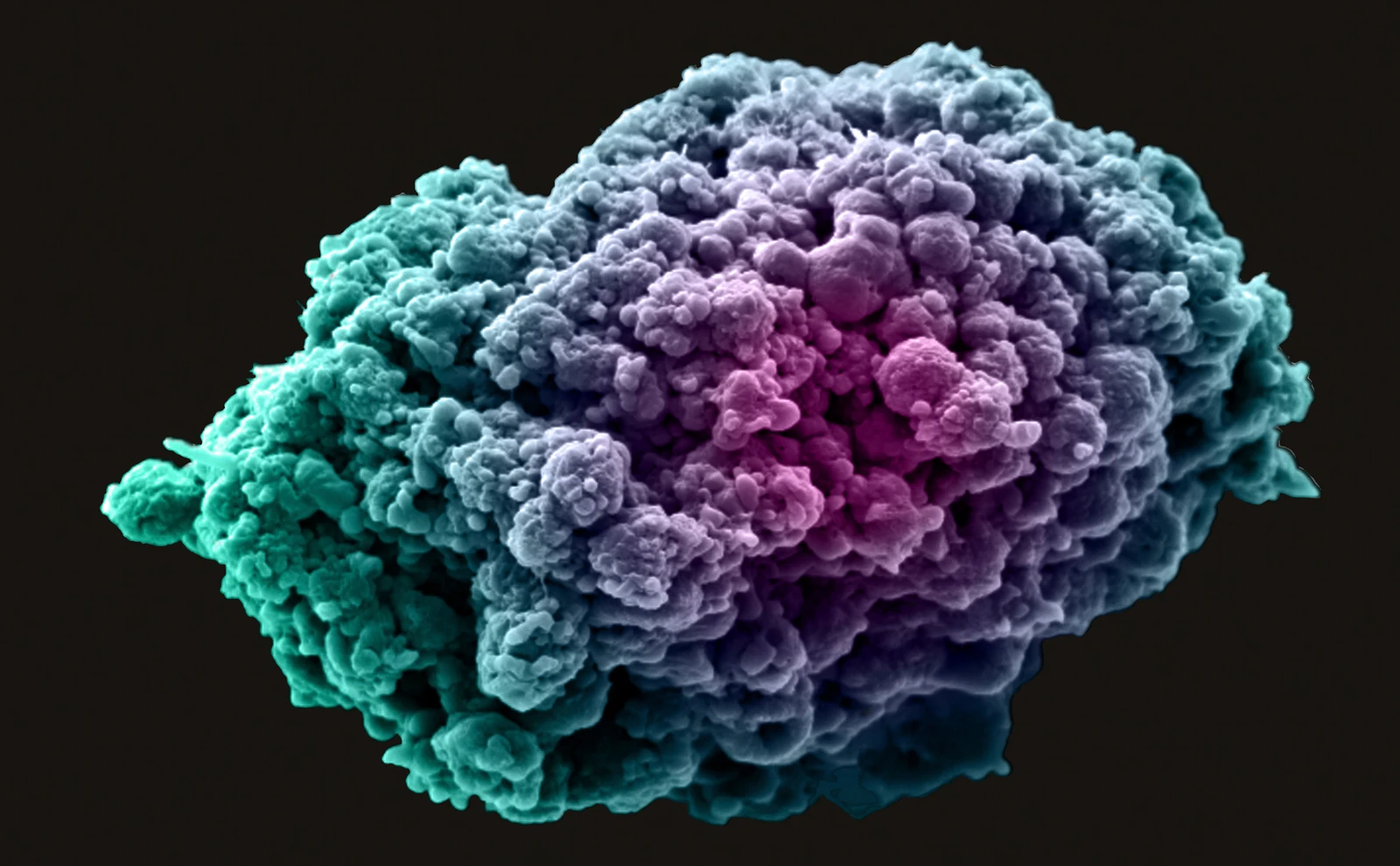
Explore whether culture setting changes a chemotherapy-associated morphological response across tumor and cardiac compartments.
Model & Design
SKBR3 breast-cancer and AC16 cardiac cell-line tissues in separate chambers; U87 glioblastoma tissue tested separately. Transwell monocultures and cocultures are comparators.
Experimental Format
Collagen at 2 mg/mL and 200,000 cells/mL; imaging on days 3 and 7. Doxorubicin 10 μM for 48 hours; the separate U87 model also receives temozolomide 200 μM for 48 hours after three days of culture.
Model Components
Component
Morphology and identity
Description
HER2, α-actinin or nestin fluorescence with actin and nuclear staining; three biological specimens and five z-stacks per specimen.
Component
Image analysis
Description
AIAPS combines thresholding, erosion and watershed segmentation of two-dimensional z-stack projections to estimate cell contours and diameters. Fluorescent-bead measurements are compared with FIJI.
Measurements & Endpoints
Projected cell diameter
Cell clustering and network organization
Lineage-associated marker retention
Analysis
Interpret significant size shifts as morphological responses. Keep the paired breast–cardiac study separate from U87 and do not equate projected size with direct survival or functional cardiotoxicity.
Published Evidence
Study Sample
Three biological specimens per tissue condition, with five z-stacks per specimen. The bead comparison uses three image replicates with five images each.
Comparison
Device versus Transwell; monoculture versus coculture; untreated versus drug-treated tissue; AIAPS versus FIJI for bead sizing.
Finding
AC16 cell size decreased significantly after doxorubicin in the device but not Transwell. SKBR3 size changed across the tested culture conditions. Figure 6 reports U87 size reduction with both drugs in the device. Day-7 breast-cell sizes and same-time U87 device/Transwell comparisons were not significantly different.
Interpretation & Limits
Established cell lines and restricted doses; no patient-response validation.
AIAPS and FIJI bead estimates differed significantly despite both falling within the nominal 1–10 μm range.
Separate breast-cancer and cardiac cell-line tissues maintained in a shared microfluidic platform changed shape after doxorubicin exposure. Automated image analysis also detected responses in a separate glioblastoma model, with cell size rather than cardiac function or direct survival as the main quantitative endpoint.