Two-Stage Automated Drug Screening in Reaggregated GBM Organoids
Screen a broad oncology library at one dose, then measure concentration responses of patient-specific top hits and compare potency with systemic exposure.
Brain & Neural ModelsOrganoids
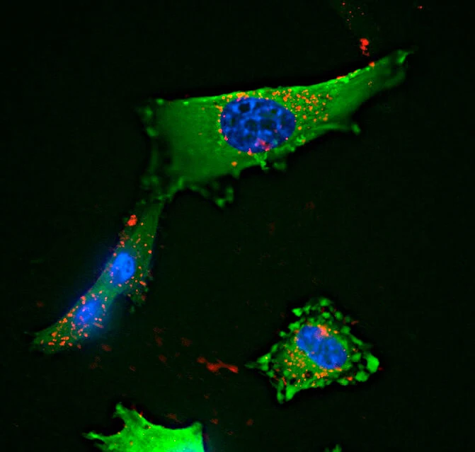
Fluorescence microscopy of cultured glioblastoma cells. (Alex Gray, CC BY 4.0)
Research Applications
Prioritize drug hypotheses in short-term surgical glioblastoma models while assessing exposure constraints.
Model & Design
Reaggregated IDH-wildtype GBM tumor organoids from viable contrast-enhancing tissue; 11 patients, nine newly diagnosed and two recurrent.
Experimental Format
25,000 cells per organoid in 384-well plates; day 2 treatment with 166 drugs at 2.5 μM for 72 hours; top five hits per case tested at 8 concentrations.
Responses are normalized to DMSO, initial hits are ranked, and dose-response curves are fitted. Serum Cmax/IC50 is a surrogate exposure comparison.
Published Evidence
Study Design
Two-stage screen and separate marizomib evaluation.
Study Sample
11 patient models in first stage; 10 with top-hit concentration-response data; seven tested with marizomib. Triplicate wells are not independent tumor repeats.
Comparison
Vehicle/untreated/positive assay controls; drug-class and patient-model comparisons.
Finding
Proteasome and selected HDAC inhibitors showed strong ex vivo activity; marizomib was weak in most tested models despite one highly sensitive case.
Interpretation & Limits
No blood–brain barrier or intratumoral pharmacokinetics in the model.
Only two recurrent cases.
Mechanistic knockdown used cell lines, not patient organoids.
A two-stage screen tested 166 approved oncology drugs in organoids from 11 glioblastomas. Selected proteasome and HDAC inhibitors showed strong ex vivo activity, while marizomib was weak in most tested models and systemic exposure remained an imperfect proxy for brain delivery.