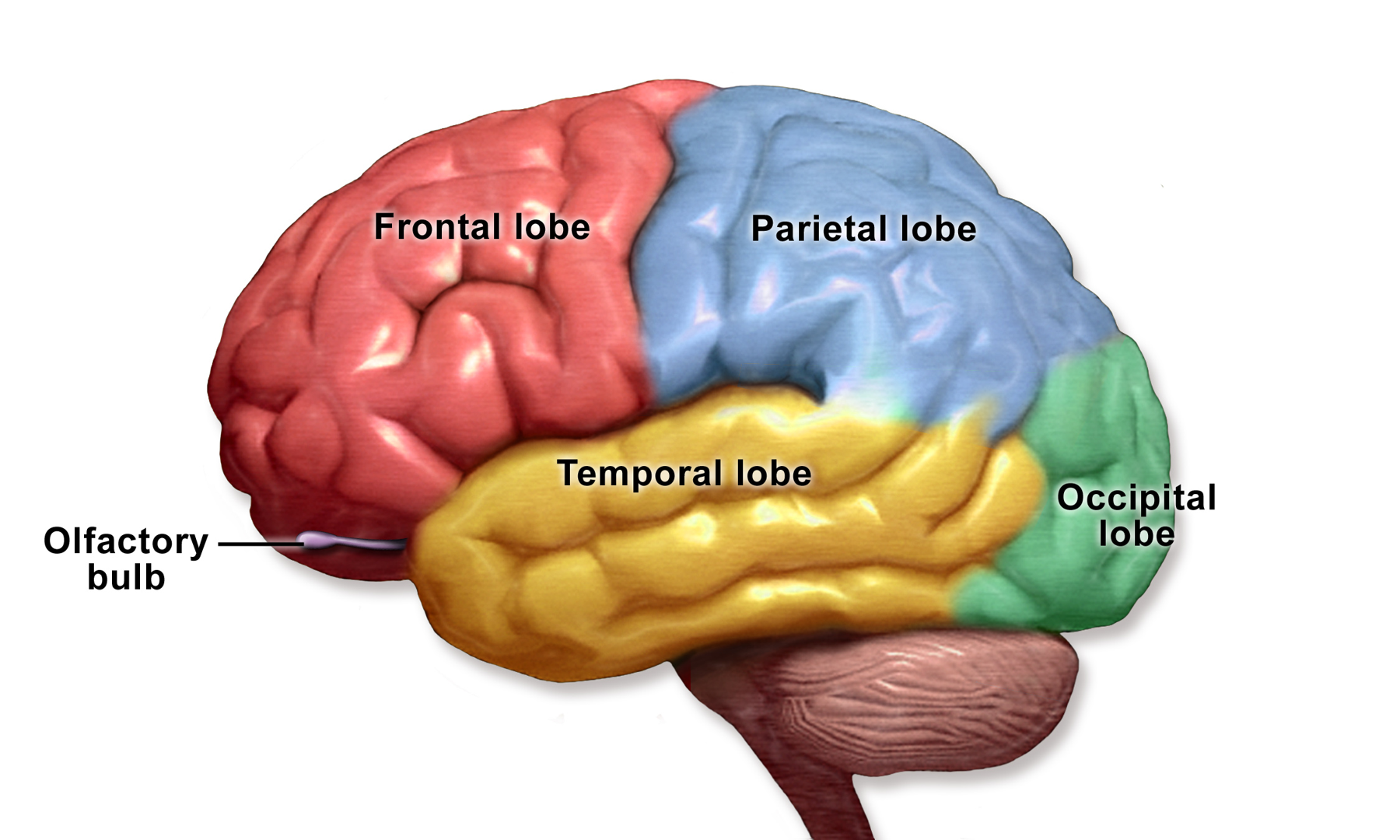
Anatomical illustration of the lobes of the human brain. (BruceBlaus / Blausen Medical Communications , CC BY 3.0 ) Research Applications Preclinical functional evaluation of an EGFRvIII-directed myeloid-cell therapeutic approach.
Model & Design Human cortical organoid-derived microglia, glioblastoma cells and combined glioblastoma–cortical organoid models.
Model Components Component Effector cells
Description Cortical organoid-derived microglia expressing an EGFRvIII-directed CAR.
Component Tumor models
Description Two-dimensional glioblastoma cocultures and three-dimensional glioblastoma–cortical organoid models.
Component Molecular analysis
Description Single-cell RNA sequencing of phagocytic pathways and internalized tumor transcripts.
Measurements & Endpoints Selective tumor-cell uptake and depletion Microglial infiltration into tumor regions Phagocytosis-associated transcriptional pathways Analysis Evaluate the 2D and 3D functional observations separately, then connect them with the single-cell measurements.
Published Evidence Study Design Two-dimensional coculture followed by three-dimensional tumor–brain organoid evaluation.
Study Sample Human cortical organoid-derived microglia and glioblastoma model systems.
Comparison Two-dimensional and three-dimensional experimental contexts.
Finding CAR microglia selectively phagocytosed tumor cells in 2D and infiltrated tumor regions with tumor depletion in 3D.
Interpretation & Limits Single-cell tumor transcripts provide molecular evidence alongside functional imaging. The study evaluates human-derived in vitro models; patient treatment responses were not established.