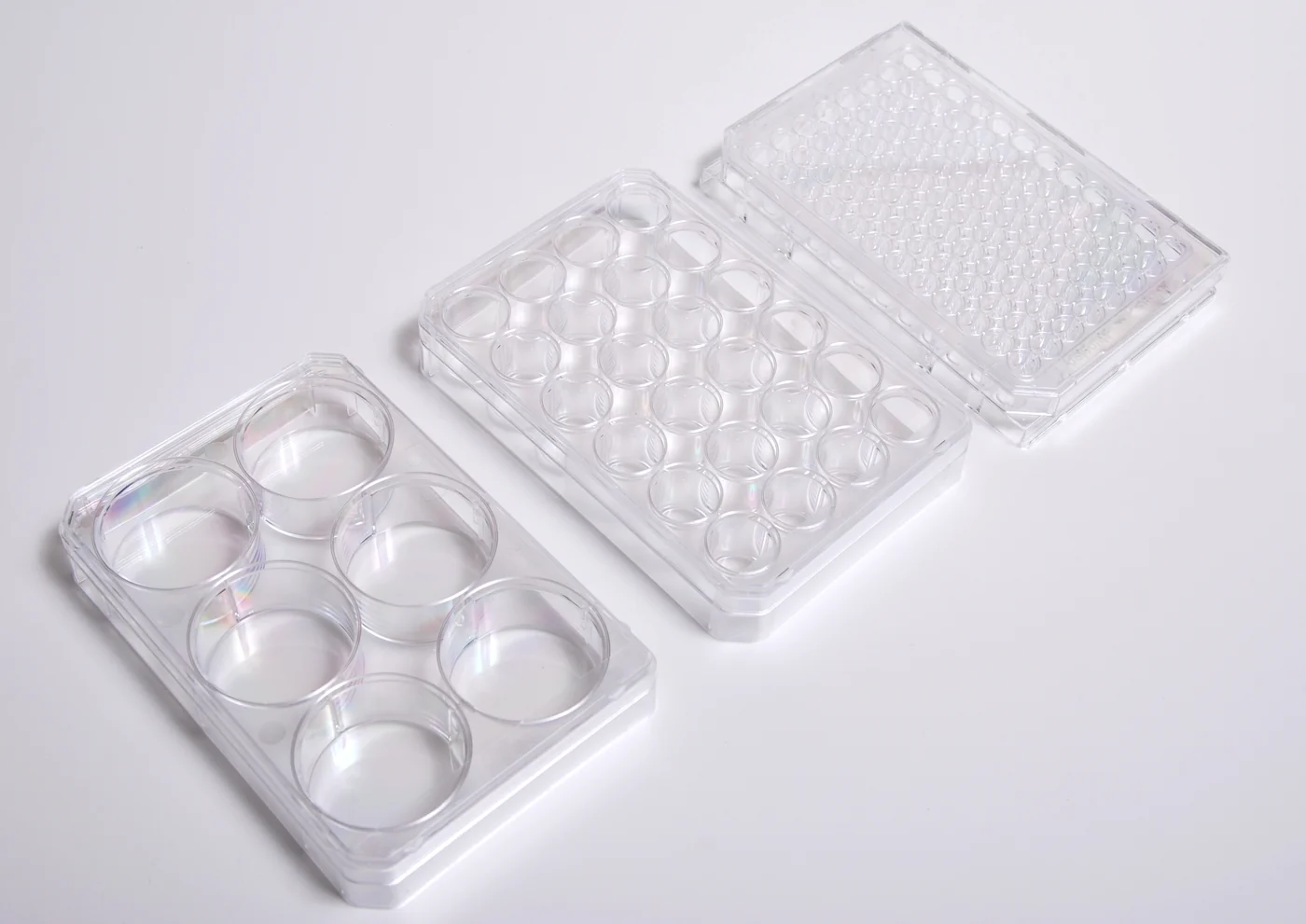
Multiwell cell-culture plates. (Lilly_M , CC BY-SA 3.0 ) Research Applications Mechanistic comparison of candidate interventions in an NPC1-associated human neuronal phenotype.
Model & Design iPSC-derived cortical neurons from one patient homozygous for NPC1 I1061T, compared with healthy-control neurons.
Model Components Component Cell systems
Description Patient and control cortical neurons expressing TUJ1/TBR1.
Component Measurements
Description Filipin fluorescence, TUNEL-positive cell proportion, NPC1 immunoblotting and morphological inspection.
Measurements & Endpoints Unesterified-cholesterol staining TUNEL-positive cell proportion NPC1 protein abundance Neuronal morphology Analysis Analyze each endpoint against its matched control and preserve the distinct concentration and time comparisons.
Published Evidence Study Design Compare patient and control baseline phenotypes, then assess JQ1 concentration and time responses.
Study Sample One patient genetic background; selected imaging/protein assays report n = 3 repeats, not three donors.
Comparison Healthy-control neurons, untreated cultures and DMSO vehicle conditions.
Finding JQ1 reduced filipin and TUNEL-associated measurements and increased NPC1 protein, with the largest responses generally at 72 hours.
Interpretation & Limits The relationship between BET inhibition, NPC1 abundance and cholesterol handling remains mechanistically unresolved. A single patient variant/background and short exposure limit population and long-term inference. The source is a preprint.