
An ALS Imaging Protocol Separates Distal Axon Damage from Muscle Background
A human motor-neuron–myotube protocol combines compartmentalized culture with Fiji and CellProfiler analysis. It measures axonal structure separately from pTDP-43 puncta restricted to intact axons, preserving the spatial meaning of each endpoint.
A distal axon needs its own measurement region
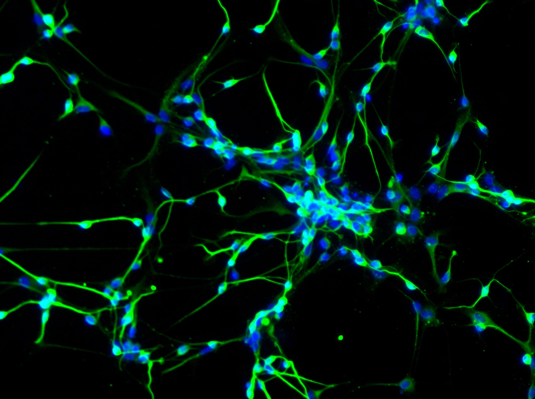
In a neuromuscular coculture, an axon may extend across a large myotube whose image contributes substantial background. Counting a protein signal across the whole field can therefore obscure where pathology lies. A Bio-protocol article from Tel Aviv University describes a human motor-neuron–myotube system and an image-analysis workflow designed specifically for distal axons in amyotrophic lateral sclerosis (ALS) models.
The model uses motor neurons and myotubes differentiated from induced pluripotent stem cells (iPSCs), with ALS genotypes and corresponding isogenic controls. A microfluidic device separates neuronal cell bodies from a distal compartment containing muscle. Axons cross connecting microgrooves, allowing the distal region to be analyzed independently. Cultures are maintained for approximately 4–6 weeks to establish the neuromuscular system and the phenotypes considered in the protocol.
The article's methodological contribution is the combination of that spatial separation with channel-specific segmentation. Neurofilament heavy chain (NFH) identifies axonal structure. Phosphorylated TDP-43 (pTDP-43), a disease-associated protein form, provides a separate molecular signal. Neither measurement is inferred solely from the overall appearance of the muscle-containing field.
Two image pipelines answer different questions
Fiji, the ImageJ-based image-processing software, is used to inspect channels, choose thresholds and create binary masks. CellProfiler, open-source software for quantitative microscopy, then identifies objects and measures their areas or counts. The protocol provides macros and analysis pipelines through the authors’ repository.
One workflow identifies intact and degenerated NFH-positive structures. Larger continuous structures are distinguished from smaller fragmented or discontinuous objects using size, shape and intensity features. This supplies the structural information for the NFH degeneration index used in the source research.
The other workflow identifies pTDP-43 puncta inside the mask of intact NFH-positive axons. Degenerated fragments are excluded from this molecular analysis. Its outputs include puncta area normalized to NFH area and puncta count normalized to NFH area. The latter density measurement is described as an additional step beyond the previously published area-based analysis.
That distinction defines the biological meaning of the result. The molecular endpoint quantifies signal within retained intact axons; it does not measure all pTDP-43 present throughout the distal compartment. The structural and molecular pipelines must therefore be interpreted together when comparing a disease phenotype or an intervention.
The publication separates fields from independent cultures
The protocol recommends systematic acquisition of distal images, avoiding repeat imaging of the same region and maintaining the same settings across conditions. It describes channel-specific parameter selection, then applying the recorded settings across the relevant dataset. The same types of segmentation parameters can be used in both pipelines without assuming identical numerical threshold values for different channels.
The representative disease comparison uses TDP-43 M337V and isogenic control cocultures. Figure 4 reports differences between the groups in axonal pTDP-43 area and the NFH degeneration index (unpaired, two-tailed t-tests; P = 0.0001). The comparison includes 28 and 18 imaging fields from 3 independently grown neuromuscular cocultures. These quantitative outcomes were adapted from the 2025 research article, while the 2026 publication documents the procedure and its analysis in greater detail.
The authors state that image datasets were blinded and independently analyzed after initial pipeline validation. They also call for reviewing segmentation overlays and excluding objects that were not properly segmented. Area conversion requires the pixel dimensions of the particular image, preserving the difference between pixel counts and physical area.
A quantitative endpoint for mechanistic and intervention studies
The earlier application examined muscle-derived miR-126 and axonal TDP-43 pathology. The protocol uses that work as its validation reference; it does not report a new drug-efficacy trial. Its relevance to therapeutic research lies in supplying compartment-specific structural and molecular endpoints that can be evaluated when an intervention is tested in the human coculture.
The authors propose that the modular analysis could be adapted to other axonal proteins or neuronal types. Such transfer requires checking the new staining, morphology and segmentation, rather than assuming the current thresholds will remain valid. The value of the workflow is the explicit relationship between region, marker and denominator, which lets a change in distal-axon structure be distinguished from a change in protein signal inside the axons that remain intact.