Compartment-Specific Axon Structure and pTDP-43 Analysis in Human Neuromuscular Cocultures
A semi-automated image pipeline distinguishes axonal fragmentation from protein puncta quantified inside intact distal axons.
Brain & Neural ModelsHuman Cell Assays
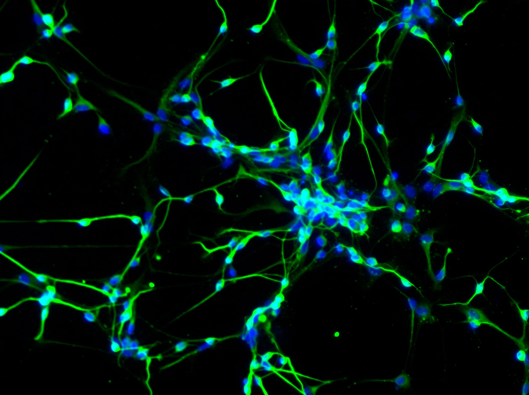
Neurons differentiated from human neural stem cells. (Yirui Sun, CC BY 4.0)
Research Applications
Supply region-specific structural and molecular endpoints for mechanistic or intervention studies in a human ALS-related coculture.
Model & Design
Human iPSC-derived motor neuron–myotube coculture with cell bodies and distal axon/muscle regions separated by microfluidic grooves.
Experimental Format
Fiji/ImageJ preprocessing and recorded channel thresholds precede CellProfiler object analysis; quantify NFH structural classes and pTDP-43 restricted to intact NFH-positive regions.
Model Components
Component
Model and markers
Description
ALS-related TDP-43 M337V and isogenic cocultures; NFH axon marker and phosphorylated TDP-43 molecular signal.
Component
Analysis resources
Description
Fiji/ImageJ preprocessing, CellProfiler pipelines, macros and overlay review; source provides a public analysis repository.
Measurements & Endpoints
Intact and degenerated NFH-positive objects
pTDP-43 area normalized to intact NFH area
pTDP-43 puncta density per NFH area
Analysis
Keep structural degeneration and molecular signal denominators distinct. The molecular endpoint excludes degenerated fragments and is not total compartmental aggregate burden.
Published Evidence
Study Design
Source protocol illustrates the analysis with disease/isogenic comparisons adapted from the 2025 validation study; post-validation image datasets were blinded and independently analyzed.
Study Sample
Figure 4: 28 and 18 imaging fields from 3 independently grown neuromuscular cocultures.
Comparison
TDP-43 M337V versus isogenic control coculture, measured within the defined distal compartment.
Finding
The M337V/isogenic-control benchmark reports differences in pTDP-43 area within intact NFH-positive axons and in the NFH degeneration index (unpaired, two-tailed t-tests; P = 0.0001). Puncta-count density is an additional pipeline endpoint beyond the earlier area-based analysis.
Interpretation & Limits
Representative outcomes derive from the 2025 source study, not a new 2026 efficacy experiment.
Only intact NFH regions enter the molecular measurement.
Image quality, segmentation and threshold values require checking for another target or neuronal type.
A human motor-neuron–myotube protocol combines compartmentalized culture with Fiji and CellProfiler analysis. It measures axonal structure separately from pTDP-43 puncta restricted to intact axons, preserving the spatial meaning of each endpoint.