Reconstructing the Choroid-Plexus Barrier
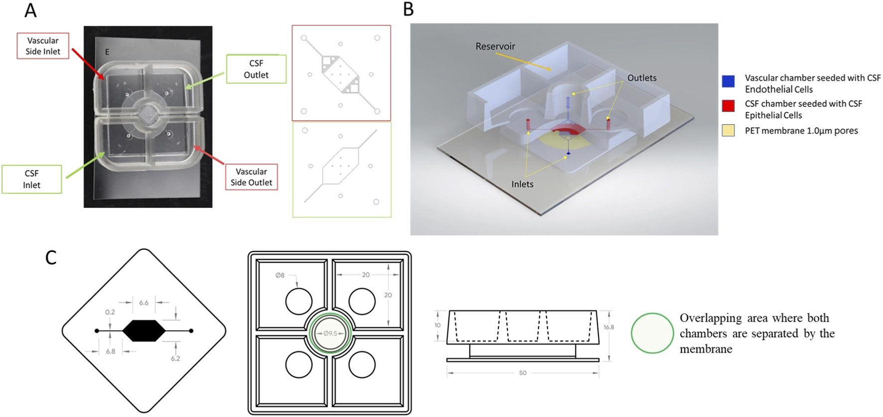
The blood–cerebrospinal fluid barrier (BCSFB), formed at the choroid plexus, is one route by which substances enter cerebrospinal fluid. Researchers at the University of Pittsburgh and Vanderbilt University, with collaborators from Eli Lilly, constructed a two-compartment microphysiological chip using commercially sourced human primary endothelial and choroid-plexus epithelial cells. A porous polyethylene terephthalate membrane separated the vascular and cerebrospinal-fluid sides, with independent medium and gravity-driven perfusion.
The team used 3 kDa fluorescent dextran transport to assess barrier function, defining an acceptable apparent permeability of no more than 1.55 × 10⁻⁵ cm/s. Across four independent experiments, 14 of 15 chips met the barrier criterion through day 15. This demonstrates a relatively sustained barrier in the tested device, while leaving between-donor and longer-term performance for further study.
This barrier differs from the endothelial blood–brain barrier. In the choroid plexus, capillaries are relatively permeable, and the epithelial layer controls exchange with cerebrospinal fluid. The chip was built around that epithelial interface, together with a vascular endothelial compartment. Keeping these cell populations in connected but distinct environments allowed the researchers to examine transport across an organized barrier while supplying the compartments with their respective culture conditions.
The team combined permeability measurements with imaging of junction proteins. ZO-1, occludin and VE-cadherin were all expressed more prominently in the epithelial layer than in the endothelial layer in this model. This characterization mattered because a low transport signal alone would not identify which cells formed the restrictive interface. The molecular images and the tracer measurements supplied different checks on the same model.
Barrier stability was also tested over time and under different ways of driving flow. In the reported operating window, gravity-driven and syringe-pump systems gave no appreciable difference in barrier performance, although neither extended the viable period indefinitely. Comparing the cellular construct with the device alone helped separate resistance created by the tissue from resistance arising from the chip. These controls are relevant to interpreting the PEG and antibiotic measurements, which otherwise could be affected by the material or fluidics as well as by the cells.
Free PEG Crossed and Accumulated Differently
The investigators then exposed the vascular side to free polyethylene glycol (PEG) preparations of different molecular weights and concentrations. Medium measurements and fluorescence imaging addressed distinct questions: how much PEG appeared in each compartment and where fluorescent signal accumulated in the cells. The 20 kDa PEG experiments showed concentration-dependent accumulation. Lower-molecular-weight PEG reached the epithelial cells more readily, whereas higher-molecular-weight PEG was associated with larger individual PEG-positive puncta in the measured images.
The dextran-based barrier measurement did not show a significant increase in tracer permeability with the PEG conditions studied. These experiments used free PEG, so they do not directly characterize transport of every PEGylated therapeutic or reproduce all exposure patterns associated with a formulated biopharmaceutical.
A Limited Drug-Transport Comparison
To test whether the device could distinguish transport of known compounds, the authors measured chloramphenicol and ceftriaxone on both sides of the barrier using mass spectrometry. Five chips were tested for each drug. Chloramphenicol showed greater transport to the cerebrospinal-fluid side than ceftriaxone (P = 0.03), in the direction expected from previously reported human observations.
That comparison is useful as an initial discrimination check, not a validation of drug-transport prediction across chemical classes. The paper's value is the combination of sustained barrier measurement, compartment sampling and cell-level imaging in a human-cell system. Further independent drugs, donor sources and quantitative comparisons would be needed to establish a wider context of use.