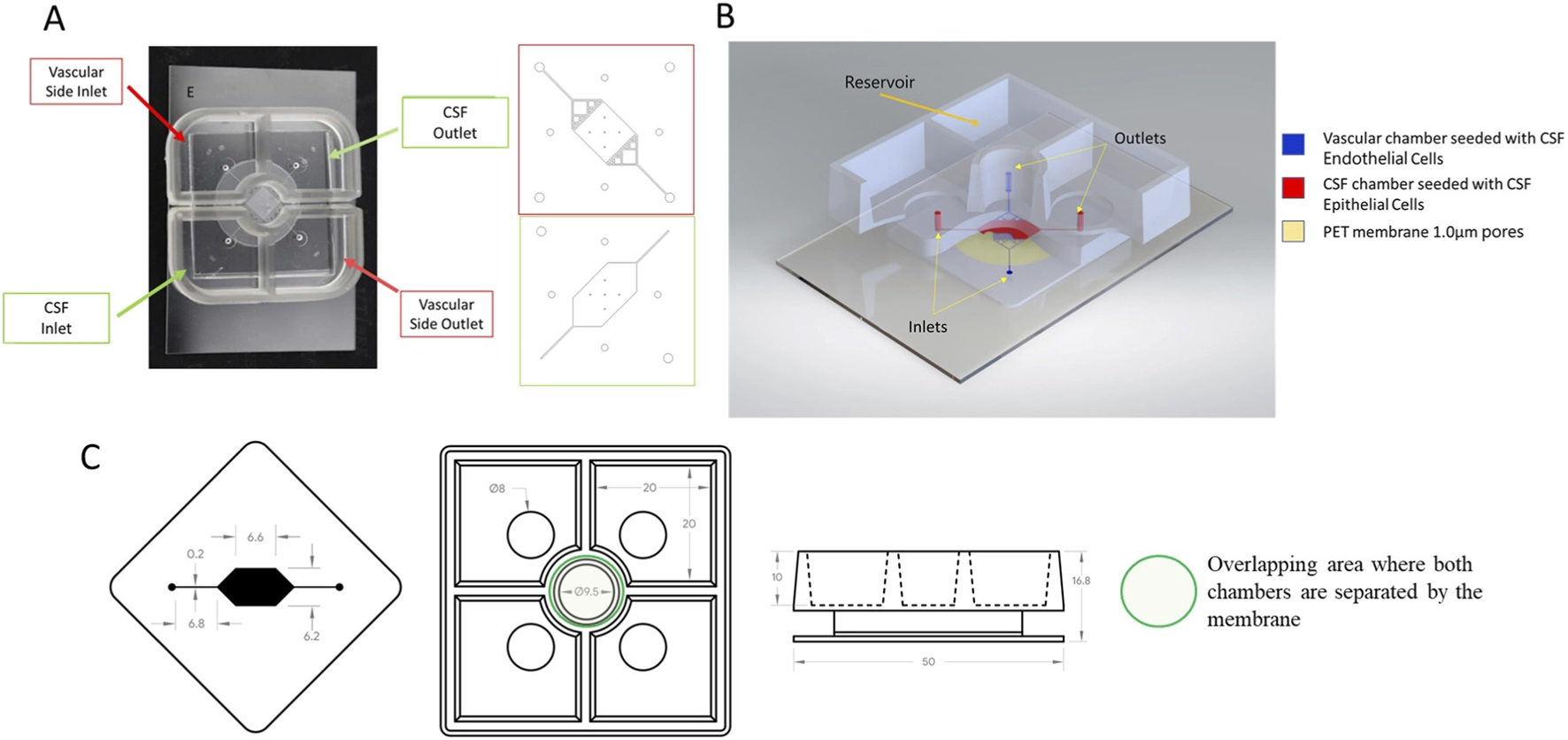
Human Blood–CSF Barrier Chip for PEG and Drug Transport
A two-compartment chip enables separate sampling on vascular and cerebrospinal-fluid sides while measuring barrier permeability, PEG distribution and drug transport.
Investigate entry into the cerebrospinal-fluid compartment, localization of free PEG and barrier changes in a defined human-cell system.
Model & Design
Commercially sourced human primary endothelial and choroid-plexus epithelial cells, tested with at least two cell batches or sources in independent experiments.
Experimental Format
A 1 μm porous PET membrane separates compartments with independent media and gravity-driven perfusion; pump perfusion was also compared.
Model Components
Component
Two-compartment chip and primary cells
Description
Endothelial cells on the vascular side and epithelial cells on the cerebrospinal-fluid side.
Component
Barrier and PEG measurements
Description
3 kDa dextran permeability, PEG immunoassay and fluorescence imaging.
Component
Drug quantification
Description
Mass-spectrometry measurements of chloramphenicol and ceftriaxone on both sides.
Measurements & Endpoints
Dextran apparent permeability
Cell viability and junction markers
PEG concentrations in each compartment
Cellular PEG puncta
Fraction of drug transported across the barrier
Analysis
Use a 3 kDa dextran permeability criterion of ≤1.55 × 10⁻⁵ cm/s. Measure medium PEG by immunoassay, cellular PEG by imaging and reference-drug transport by mass spectrometry.
Published Evidence
Study Design
Assess barrier stability under gravity perfusion, then compare free-PEG conditions and transport of two reference drugs.
Study Sample
Barrier assessment included four independent experiments and 15 chips; each drug-transport arm used five chips.
Comparison
Different PEG molecular weights and doses; chloramphenicol versus ceftriaxone, interpreted alongside published human observations.
Finding
Fourteen of 15 chips retained barrier function to day 15. PEG localization varied with molecular weight and concentration without a significant change in dextran permeability under tested conditions. Chloramphenicol crossed more readily than ceftriaxone (P = 0.03).
Interpretation & Limits
Only two reference drugs were compared; broad prediction of drug transport was not established.
PEG studies used free PEG, not all PEGylated medicines.
Longer-term performance and donor-related variation need further testing.
A two-compartment chip assembled from human primary cells maintained a measurable blood–cerebrospinal fluid barrier for up to 15 days. Its first applications compared the distribution of free PEG species and transport of two reference drugs; the latter comparison does not yet establish broad predictive performance.