
Preprint Measures Drug-Sensitive Evoked Responses in Human Cortical Organoids
Brainopheno combines electrical stimulation, microelectrode recordings and an offline decoder to measure response separability in human cortical organoids. Neural inhibitors reduced classification accuracy, while disease and monocyte comparisons revealed changes missed by some spontaneous-activity measures.
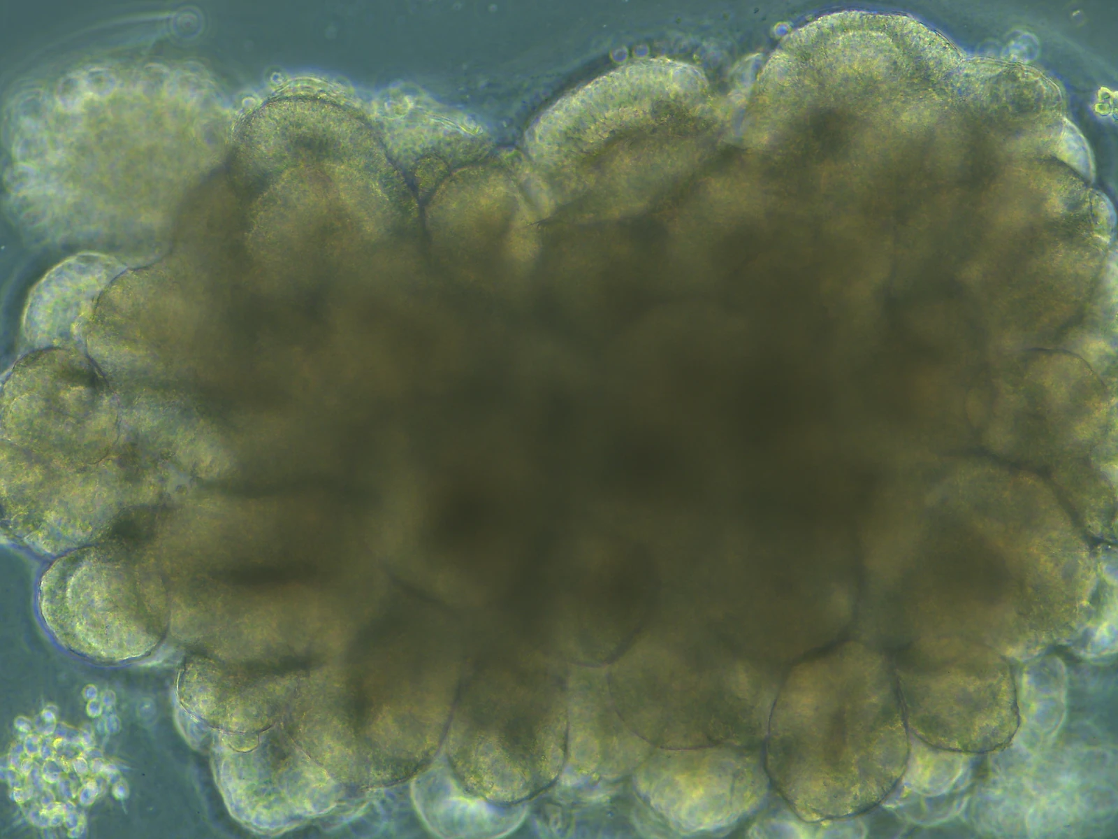
A Functional Readout Based on Evoked Activity
A bioRxiv version-one preprint posted September 10, 2026, describes Brainopheno, an assay of electrically evoked activity in human cortical organoids. Researchers led by Indiana University use a microelectrode array, or MEA, to stimulate and record the organoid surface, then apply an offline classifier to distinguish the resulting activity patterns.
The measured outcome is the separability of neural responses to different stimulation patterns. Classification is performed by a logistic-regression decoder on the recorded signals. This provides a quantitative network phenotype for pharmacological and disease comparisons; the experiment does not test an organoid’s cognition or task performance in a person.
Three Stimulation Patterns and Repeated Recordings
The reported pattern-classification experiments used three spatial stimulation patterns, labeled α, β and γ, with 30 trials per pattern. Analysis selected the 20 most active channels, excluded the first 10 milliseconds to reduce stimulus artifact and represented evoked spikes in ten-millisecond bins. A two-to-one training/test split evaluated the decoder within the recording dataset.
The strongest representations appeared around 30 milliseconds after stimulation. Pattern dissimilarity and classification accuracy increased as organoids matured from two to four months, alongside changes in functional connectivity. These comparisons link the readout to development of the cultured network. The recording-level split does not establish prediction in an independently held-out donor or organoid.
The cortical preparations contained excitatory and inhibitory neurons, progenitors and astrocytes. The source describes embryonic-stem-cell and induced-pluripotent-stem-cell (iPSC) resources, with patient and healthy-donor iPSC lines used for the Alzheimer’s comparison. Their developmental state and surface contact with the array shape which network activity the assay can sample.
Neural Inhibitors Changed the Evoked Phenotype
After twenty-minute pharmacological exposures, AP5 plus CNQX, antagonists of NMDA and AMPA/kainate receptors, reduced classification accuracy from 89% to 55.5%. Neural activity and functional connectivity also declined. The reported concentrations were 25 μM for each antagonist.
Bicuculline at 20 μM increased firing while accuracy fell from 88.5% to 78%, with comparatively little change in connectivity. Tetrodotoxin at 1 μM suppressed activity and connectivity and reduced accuracy from 85.5% to 32.5%, close to the chance level for three classes. Figure 3 reports five organoids across three independent experiments.
These perturbations show that higher firing does not necessarily produce better response separation. Combining the decoder output with spontaneous activity and connectivity measures helps distinguish mechanisms that would be blurred by a firing-rate endpoint alone. The preprint gives inconsistent ages for the pharmacological subset in Results and Methods, so the exact developmental age of that comparison remains unclear.
Disease and Immune Comparisons Added Different Signals
Three-month organoids derived from a familial-Alzheimer’s patient line carrying an APP duplication accumulated more amyloid-β than healthy-donor controls. Mean firing rate and global efficiency were similar between groups, whereas classification accuracy and pattern dissimilarity were lower in the APP organoids. The functional comparison reports 15 organoids across three experiments; amyloid quantification used six organoids.
The researchers also added circulating monocytes from young donors, older age-matched controls or Alzheimer’s patients to healthy cortical organoids. Young-donor monocytes improved the measured phenotype, older-control monocytes showed a smaller improvement, and Alzheimer’s-associated monocytes impaired it. The comparison reports 14 organoids across three experiments. Sequencing provided associated neuronal transcriptional changes rather than direct proof of a single causal immune pathway.
Extending the Assay Beyond Proof of Concept
The study proposes evoked-response phenotyping as an additional endpoint for neural drug and disease models. Surface electrodes sample a restricted portion of a three-dimensional organoid, while tissue heterogeneity and diffusion limit reproducibility and access by compounds or immune cells. The authors also selected organoids with adequate early activity for the monocyte experiments. Wider validation will need to establish how those selection criteria, donor background and device geometry affect response ranges before the assay is used to compare candidate therapies.