
Preprint Tests Halofantrine in Mouse and Patient-Derived Retinal Organoids
A staged screen identified halofantrine as a candidate photoreceptor protectant. Human follow-up used a CEP290-associated retinal degeneration line and an unaffected family control; animal studies added functional signals, route-dependent exposure and unresolved dose effects.
A Mouse Screen Led to a Human Disease Model
An NIH-led Research Square preprint investigates halofantrine, a compound previously developed as an antimalarial, as a candidate treatment for retinal degeneration. The May 13, 2026 version combines a mouse retinal organoid screen, a patient-derived human organoid comparison and follow-up experiments in rodents. These stages address different questions: candidate selection, preservation of human disease-associated photoreceptor features and effects on retinal function and tissue exposure.
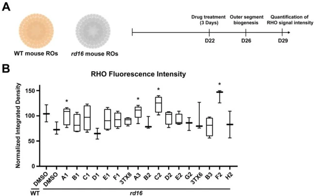
The initial screen tested 17 molecules in retinal organoids from Cep290 rd16 mutant mice. Compounds were given for three days beginning at differentiation day 22, with rhodopsin fluorescence assessed at day 29. Halofantrine, designated A3, was selected alongside other candidates for further testing. Rhodopsin signal provided a rod-photoreceptor readout; it did not measure visual function in the organoids.
For human follow-up, the researchers used induced pluripotent stem cells from one child with compound-heterozygous CEP290-associated Leber congenital amaurosis and the child’s unaffected mother. Treatment began at organoid day 90, before the later emergence of prominent photoreceptor abnormalities. Reserpine was tested at 30 μM, while halofantrine and two other candidates were tested at 10 μM. The reported imaging experiments included at least three independent groups, with at least four organoids per group.
Human Organoids Distinguished Rescue, Toxicity and Unsuccessful Combinations
Both reserpine and halofantrine significantly increased rhodopsin staining in the affected human organoids. Another candidate, F2, showed a favorable trend that did not reach significance. Candidate C2 caused cellular toxicity and was excluded. Halofantrine also partly improved the ARL13B ciliary phenotype. The authors did not observe adverse changes in the reported cone, bipolar-cell or synaptic staining under the tested conditions.
Combining lower-dose halofantrine with reserpine or F2 did not further increase the response relative to halofantrine alone. The human follow-up describes a single CEP290 family background.
Animal Follow-Up Produced Several Different Functional Outcomes
In female P23H rats, which model a separate inherited retinal degeneration, halofantrine produced the most persistent benefit among the candidates in the optokinetic tracking test. Electroretinography also indicated preservation of b-wave responses, while a-wave improvements were not significant. The vehicle group contained seven rats and compound groups contained six or seven. Histology supported preservation of rod-associated markers, outer nuclear layer structure and cone staining.
In rd10 mice carrying a Pde6b mutation, halofantrine improved the photopic b-wave response. The scotopic b-wave showed a nonsignificant trend, and a-wave changes were not significant. Tissue measurements again supported photoreceptor preservation. The agreement across some structural and functional endpoints strengthens the candidate signal, while the different electrophysiological outcomes prevent a claim of uniformly restored retinal function.
Transcriptomic comparisons identified 46 differentially expressed genes shared by at least two models. ITPR3 changed in all three, and oxidative phosphorylation, ribosomal and circadian-entrainment pathways appeared across the models. These associations suggest processes for mechanistic follow-up; they do not identify the molecular target responsible for protection.
Local Delivery Changed Exposure and Did Not Give a Simple Dose Response
The preprint separately compared ocular routes and concentrations. In P23H rats, 25 μM subconjunctival halofantrine improved the reported later optokinetic and b-wave outcomes. Intravitreal 50 μM initially suppressed a scotopic b-wave response before recovery, whereas 100 μM did not show that initial suppression. Several other route-comparison endpoints, including a-waves, remained nonsignificant. The response therefore did not follow a straightforward concentration ordering.
Pharmacokinetic experiments in wild-type animals found route-dependent retinal and surrounding-tissue exposure. Suprachoroidal delivery gave an early retinal peak, intravitreal delivery showed more persistent retinal levels, and subconjunctival delivery favored surrounding ocular tissues. Plasma concentrations were below the 1 ng/mL quantification limit in the measured samples. That analytical limit does not establish absence of systemic exposure or resolution of halofantrine’s known cardiac liability. Poor water solubility, nonuniform dose effects and the need to connect ocular exposure with durable benefit remain central development questions. Eyedrop delivery was proposed rather than tested.