Staged Retinal Organoid Screening and Human CEP290 Candidate Retesting
Preprint method combining a 17-compound mouse retinal organoid screen with selected drug tests in one affected human CEP290 family model.
RetinaOrganoids
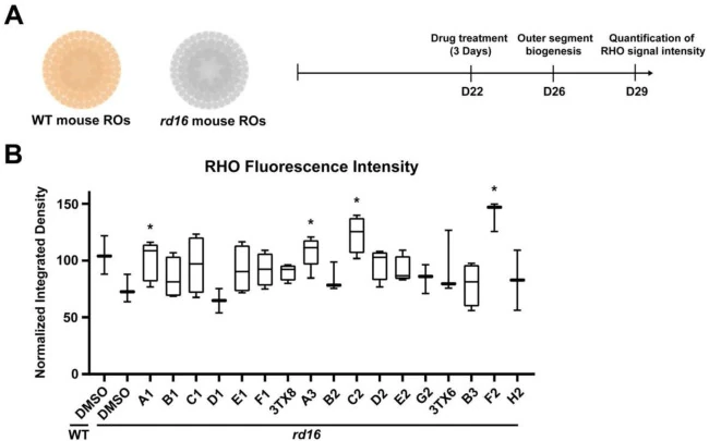
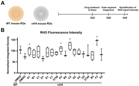
Preprint Figure 1A–B: mouse retinal organoid treatment timing and rhodopsin fluorescence in the 17-candidate screen. A3 denotes halofantrine. (Kim M et al., CC BY 4.0)
Research Applications
Prioritize compounds that preserve photoreceptor-associated features, then compare response, toxicity and unsuccessful combinations in human disease-associated organoids.
Model & Design
Cep290 rd16 mouse retinal organoids for discovery; human iPSC-derived retinal organoids from one affected compound-heterozygous CEP290 child and the unaffected mother for follow-up.
Experimental Format
Three-day mouse treatment beginning at day 22 with day 29 rhodopsin staining; human treatment beginning at day 90 with immunofluorescent rod, ciliary and other retinal markers.
Model Components
Component
Discovery and human exposures
Description
17 discovery compounds. Human reserpine 30 μM; halofantrine (A3), C2 and F2 each 10 μM. Selected lower-dose halofantrine combinations were also compared.
Component
Readouts and controls
Description
Rhodopsin fluorescence, ARL13B cilia staining and cone/bipolar/synaptic markers; vehicle and unaffected-family controls. Animal functional and exposure follow-up are separate validation arms.
Measurements & Endpoints
Rhodopsin fluorescence intensity
ARL13B-associated ciliary phenotype
Candidate toxicity and combination response
Separate rodent ERG, optokinetic and ocular-exposure measurements
Analysis
Matched disease and family-control organoids are compared under the same treatments. Organoid and experimental-group counts describe replication within one affected family background. Rod-marker preservation in organoids and visual-function measurements in animals are separate endpoints.
Published Evidence
Study Design
Sequential mouse discovery and human disease-associated retesting, followed by separate retinal-degeneration animal models.
Study Sample
One affected human donor and one unaffected mother; Figure 2 reports at least three independent groups with at least four organoids per group.
Comparison
Vehicle, reserpine, other prioritized candidates, unaffected-family organoids and lower-dose combinations.
Finding
Reserpine and halofantrine significantly increased human rhodopsin staining. F2 had a nonsignificant trend; C2 was toxic. Tested halofantrine combinations added no further augmentation. Ciliary rescue was partial.
Interpretation & Limits
Preprint findings from one affected family background; the human organoid harvest time is not stated in the reported narrative.
Rhodopsin and ciliary signals are distinct from the visual-function measurements made in animals.
Animal a-wave and several b-wave/route outcomes were nonsignificant, and local delivery produced nonmonotonic dose effects.
Plasma below the 1 ng/mL quantification limit leaves low systemic exposure unresolved and does not establish cardiac safety.
A staged screen identified halofantrine as a candidate photoreceptor protectant. Human follow-up used a CEP290-associated retinal degeneration line and an unaffected family control; animal studies added functional signals, route-dependent exposure and unresolved dose effects.