
A Cardiac-Model Review Connects Tissue Design with Drug-Evaluation Endpoints
A review compares engineered heart tissues, chamber models, cardiac organoids and microphysiological systems. It organizes drug evaluation around structural, electrical, mechanical, molecular and metabolic evidence, while examining maturity and exposure constraints.
Different Cardiac Platforms Answer Different Questions
A review by researchers at Sejong University, Georgia Tech and Emory University examines human in vitro cardiac models for drug evaluation. It follows the field from single cells and aligned monolayers to engineered tissues, chamber-forming constructs, organoids and microphysiological systems. The comparison links each architecture with the biological features and measurements it makes accessible.
Human induced pluripotent stem cell-derived cardiomyocytes are central to many of these platforms. They provide a human genetic background and can be combined with supporting cell types, extracellular matrices and defined stimulation. The resulting tissues differ in maturation, lineage composition, mechanical organization and the degree of environmental control. Those differences shape their immediate drug-research use.
Monolayer and single-cell systems offer direct access to electrical and calcium-related behavior. Engineered heart tissues add aligned three-dimensional muscle and mechanical constraints, enabling force-related measurements. Chamber models extend that design to pumping geometry, pressure–volume relationships and responses to loading. The review treats these features as distinct experimental capabilities rather than a single scale of model quality.
Developmental Organization and Controlled Tissue Function
Cardiac organoids use self-organization to reproduce selected aspects of early heart development. The review describes models with cavities, epicardial components, multiple cardiac lineages and chamber-associated identities. These features support questions involving developmental signaling, tissue patterning and interactions between lineages.
Their architecture also depends on differentiation and self-organization, which introduce variability. Compared with an externally constrained engineered tissue, an organoid may provide less direct control over force transmission or tissue geometry. The developmental stage is therefore part of the intended application, especially when using these preparations to study a process characteristic of the adult heart.
Heart-on-chip systems introduce additional control over flow, electrical pacing, mechanical stimulation and the surrounding chemical environment. Depending on the design, they can support repeated measurement of contraction, conduction and injury during exposure. Incorporating vascular or immune components creates further ways to investigate inflammatory and endothelial contributions to cardiac responses.
Characterizing Structure and Function Together
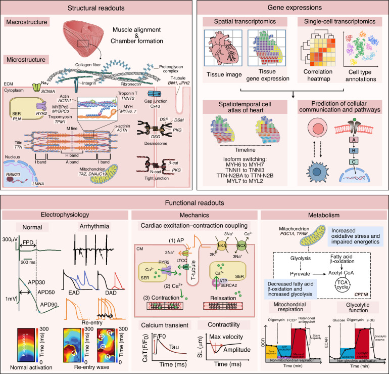
The review organizes model assessment into structural, functional, molecular and metabolic dimensions. Structural measurements include fiber alignment, sarcomere organization, cell junctions, extracellular matrix and chamber shape. Imaging can resolve these features at tissue, cellular and subcellular scales.
Functional assessment follows excitation–contraction coupling. Patch-clamp recordings and multielectrode arrays examine electrical activity; calcium imaging follows intracellular signals; cantilevers, muscular thin films and related mechanical systems measure contraction. Together, these approaches characterize electrical activity, calcium handling and mechanical performance.
Molecular profiling supplies another level of characterization. Bulk and single-cell RNA sequencing describe identity, heterogeneity and disease-associated programs, while spatial approaches retain information about where those programs occur. Protein and epigenomic measurements can complement the transcript data.
Metabolic characterization includes oxygen consumption, glycolytic activity, mitochondrial properties and substrate use. These measurements matter because hiPSC-derived cardiomyocytes often retain a relatively immature metabolic state. A preparation may have organized sarcomeres and visible beating while still differing from adult myocardium in calcium handling or energy metabolism.
Adding Exposure, Other Organs and Longer Time Scales
For drug evaluation, cardiac tissue behavior also depends on the exposure delivered to it. The review discusses liver–heart and other multi-organ configurations that connect metabolism and distribution with cardiac outcomes. These systems can examine how another tissue changes a drug’s concentration or contributes a metabolite before the cardiac response is interpreted.
Such integration introduces its own design requirements. Tissue-specific media, flow rates, relative tissue amounts and communication pathways need to remain compatible with the question. The review identifies the lack of standardized multi-organ arrangements as a constraint on cross-study comparison.
Time is another major variable. Many assays measure acute electrophysiological, contractile or viability changes over hours or days. Cumulative structural injury, fibrosis-associated remodeling and long-term functional decline require stable tissue behavior during repeated or prolonged exposure. The authors describe advances in extended culture while identifying chronic drug-response prediction as an area requiring further evidence.
Building Evidence for a Defined Drug-Research Use
The review emphasizes limitations arising from immature cell states, incomplete vasculature and differences between donors, lines and laboratories. Adding more cell types can introduce additional variation because their maturation and culture requirements differ. Larger tissues can also develop transport limitations that change their metabolic state or viability.
Reference compounds and clinically anchored comparisons help determine how a particular assay performs. The review discusses cardiac electrophysiology frameworks, including CiPA and ICH E14/S7B, while distinguishing the evidence for selected ion-channel-related effects from less established prediction of chronic structural injury.
The resulting framework begins with the drug-research question and the biological response that needs to be measured. Model architecture, maturity, exposure control and the combination of endpoints can then be selected to address it. The review organizes these capabilities and evidence requirements for defined drug-research uses.