Mapping a Human Tissue Model
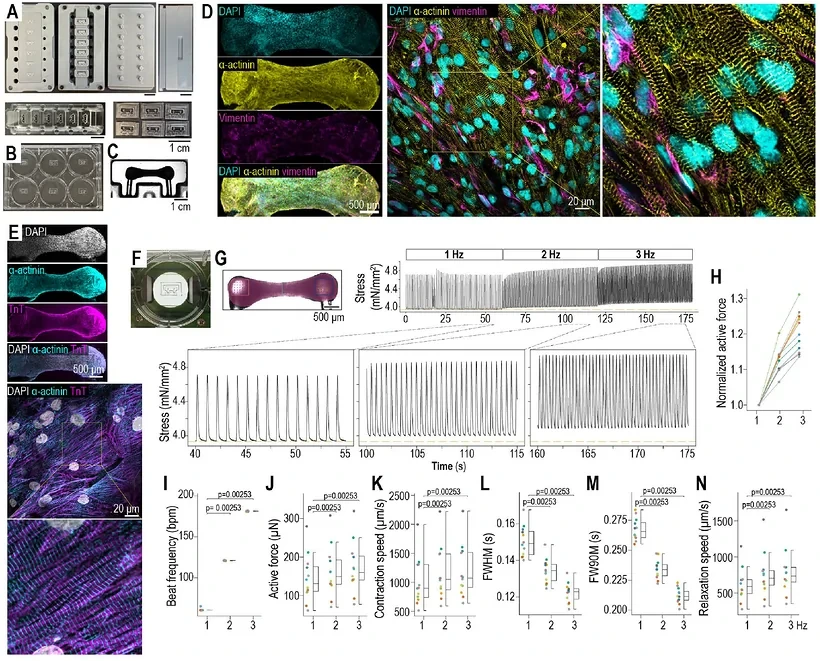
A study from Houston Methodist Research Institute and collaborators combines human engineered heart tissues (EHTs) with simultaneous voltage and calcium mapping to investigate ventricular tachycardia mechanisms. Cardiomyocytes derived from two human induced pluripotent stem cell lines were combined with primary human ventricular fibroblasts at a 3:1 ratio in collagen hydrogel. Modified milliPillar modules supported the tissues during culture and subsequent recording.
At about one month, the tissues displayed organized contraction and rate-dependent electrical responses. Dual-channel optical mapping, using RH-237 for voltage and Rhod-2 AM for calcium, achieved 1-ms temporal and approximately 22-μm spatial resolution. This allowed the investigators to relate local repolarization changes to conduction block, wavebreak and rotating activation. Action-potential duration at some pacing conditions fell within adult-human reference ranges, while longitudinal conduction remained about half the reported adult value.
A Drug Response and a Proarrhythmic Challenge
The initial pharmacological validation used 100 nM E-4031, a selective hERG blocker. At 2-Hz pacing, exposure reproducibly prolonged action-potential duration at 80% repolarization, APD80. The modest change in activation time was not statistically significant.
The investigators then combined 500 nM E-4031 with reduced potassium and magnesium to model acquired long-QT conditions. The principal mapping comparison included 27 control and 28 treated tissues from the two cell lines across six batches. In a separate sequential comparison of six tissues, neither baseline solution nor reduced electrolytes alone produced rotors; all six showed rotors under the combined condition. This provides a within-tissue comparison of baseline, electrolyte-reduced and combined-challenge conditions.
Cellular Triggers Did Not Always Become Reentry
Early afterdepolarizations occurred in 25 of 28 long-QT-condition tissues. Mapping distinguished local events, partially propagating depolarizations and triggered beats that spread through the tissue. Their relationship to calcium activity and regional conduction could be examined within individual recordings.
Phase singularities and rotors were found in 16 of 28 treated tissues and none of 27 controls. They accompanied spatially heterogeneous repolarization, reduced upstroke velocity, conduction block and wavebreak. Across 111 detected events, median duration was approximately 0.4 seconds, with activity concentrated near the tissue heads and necks. The difference between the frequency of afterdepolarizations and reentry indicates that a cellular trigger also required permissive local propagation conditions.
A minority of tissues developed more complex, fragmented activation resembling ventricular fibrillation, usually returning quickly to a more organized tachycardia-like pattern. The tissue geometry supported these transient mechanisms while placing limits on sustained disorganization.
An Independent Test of a Fixed Structural Obstacle
A separate cohort of ten tissues was examined before and within five minutes after focal cryoinjury in standard physiological solution. These experiments used no long-QT electrolyte or pharmacological challenge, and each tissue served as its own pre-injury control.
Localized damage appeared in the targeted head in all ten tissues. Phase singularities were detected in seven of ten tissues after injury, compared with one of ten beforehand. Rotor trajectories preferentially followed the border between injured and intact tissue. Three rotors lasted at least ten seconds, and some remained present when recording stopped. Their measured durations are consequently lower bounds set by the observation window.
The injury experiment complements the drug–electrolyte condition: a fixed obstacle could anchor more persistent reentry on the same basic tissue geometry in which functional rotors were generally brief.
Scope for Drug-Response Research
The study establishes pharmacological sensitivity and provides a human platform for examining how cellular triggers interact with tissue-scale propagation. E-4031 was used to validate hERG sensitivity and construct a proarrhythmic challenge; antiarrhythmic-treatment efficacy was not tested.
The authors identify several limits. Two cell lines do not cover broad genetic variability, and the tissues retain an intermediate electrophysiological state. Focal cryoinjury models the acute consequences of localized cell loss without ischemia–reperfusion, inflammatory infiltration or chronic fibrotic remodeling. Mapping is weighted toward the upper tissue surface, dyes and electromechanical uncoupling can influence electrophysiology, and short recording windows constrain assessment of long-term rotor stability.