Dual Optical Mapping of Triggered and Injury-Anchored Arrhythmias in EHTs
Simultaneous voltage and calcium mapping compares hERG/electrolyte perturbation with focal injury in engineered human cardiomyocyte–fibroblast tissues.
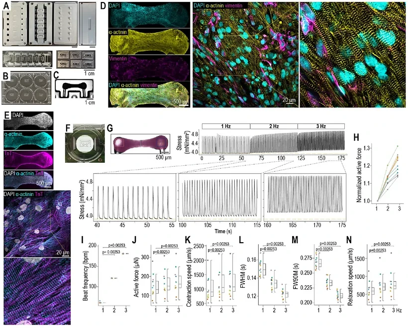
Two human induced pluripotent stem cell-derived cardiomyocyte sources combined with primary human ventricular fibroblasts at a 3:1 ratio in collagen.
Experimental Format
Approximately one-month-old milliPillar tissues; RH-237/Rhod-2 AM mapping at 1 ms and approximately 22 μm resolution.
Model Components
Component
Engineered cardiac tissue
Description
Two cardiomyocyte sources and primary human ventricular fibroblasts in a collagen tissue anchored at both ends.
Component
Pharmacological/electrolyte conditions
Description
100 nM E-4031 tests sensitivity; 500 nM E-4031 with 2 mM potassium and 0.5 mM magnesium produces acquired-long-QT-like conditions.
Component
Focal structural perturbation
Description
A separate cohort is recorded before and within five minutes after focal cryoinjury in standard physiological solution.
Measurements & Endpoints
APD80, conduction velocity and repolarization heterogeneity
Voltage–calcium activity and early afterdepolarizations
Triggered beats, conduction block and wavebreak
Phase-singularity/rotor occurrence, location and duration
Analysis
Combine regional voltage/calcium traces, activation/repolarization maps and phase analysis. Analyze the pharmacological and paired-injury cohorts separately and interpret durations within the observation window.
Published Evidence
Study Design
Compare long-QT-like challenge with controls and examine the combined perturbation by sequential exposure.
Study Sample
27 control and 28 treated tissues across six batches; a separate sequential comparison uses six tissues.
Comparison
Control versus combined challenge; sequential baseline, reduced electrolytes and reduced electrolytes plus E-4031.
Finding
Phase singularities/rotors occurred in 16/28 versus 0/27 tissues; sequential counts were 0/6, 0/6 and 6/6. Median duration across 111 events was approximately 0.4 s.
Interpretation & Limits
The 100 nM sensitivity experiment differs from the 500 nM/electrolyte challenge.
Antiarrhythmic-treatment efficacy was not tested.
Study Design
An independent cohort is compared before and after focal injury.
Study Sample
Ten tissues from two hiPSC-cardiomyocyte sources, each serving as its own pre-injury control.
Comparison
Standard physiological solution before versus after focal cryoinjury, without the long-QT pharmacological challenge.
Finding
Phase singularities occurred in 7/10 after injury versus 1/10 before. Tracks favored the injury boundary; three rotors persisted for at least 10 s, with some ongoing at recording end.
Interpretation & Limits
An acute localized-cell-loss model without chronic inflammation/fibrosis or ischemia–reperfusion.
Two lines, surface-weighted mapping and short recordings limit extrapolation; ongoing-event durations are lower bounds.
Dual voltage–calcium mapping reveals short-lived reentry under pharmacological long-QT conditions and a subset of more persistent, injury-anchored rotors in human engineered heart tissues.