
Injured Kidney Organoids Alter Cardiac Microtissue Function in Co-culture
Human kidney organoids were injured before co-culture with cardiac microtissues. Doxorubicin-injured kidneys produced stronger secondary cardiac changes than the gentamicin condition, with distinct effects on viability, contraction and endothelial structures.
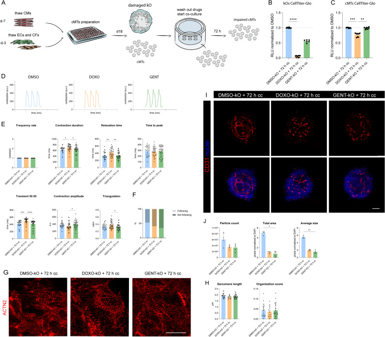
Separating Kidney Injury from the Cardiac Observation
A Leiden University Medical Center-led team combined human iPSC-derived kidney organoids with cardiac microtissues to study how an injured renal tissue can affect the heart in vitro. The design separates the initial drug exposure from the subsequent organ interaction: kidney organoids are treated first, washed and then placed in co-culture with cardiac tissues.
The kidney cultures received either 2 μM doxorubicin or 1 mg/mL gentamicin for 72 hours. Both conditions reduced renal viability and altered tissue morphology and injury-associated expression. The organoids showed changes in glomerular and tubular structures, providing defined injured tissues for the next stage of the experiment.
Cardiac microtissues contained cardiomyocytes, fibroblasts and endothelial cells. They were co-cultured for a further 72 hours with either injured or control kidney organoids in a static Transwell arrangement. The model therefore permits soluble interactions between separated tissues while retaining each tissue for its own measurements.
Different Kidney Exposures Produce Different Cardiac Responses
Cardiac viability decreased after co-culture with doxorubicin-injured kidney organoids. The gentamicin-injury condition produced a weaker response and did not significantly reduce cardiac viability in the corresponding comparison. These results show that the secondary cardiac phenotype depends on the state of the injured kidney tissue, rather than appearing uniformly after any renal insult.
The investigators also measured contraction under 1 Hz pacing. A greater proportion of cardiac microtissues failed to follow the pacing stimulus after co-culture with injured kidneys. Contraction parameters were then calculated for tissues that did respond, with the nonresponding fraction retained as a separate result.
Among responding tissues, the doxorubicin-kidney condition prolonged contraction duration, relaxation and the reported 50–50 transient duration. Time to peak and contraction amplitude were not significantly changed. Sarcomere measurements were relatively preserved, while CD31-positive endothelial structures declined. The combined observations point to functional and vascular changes without uniform loss of every structural feature.
Examining Each Tissue's Transcriptional Response
Bulk RNA sequencing was performed separately on the kidney and cardiac tissues after co-culture. The doxorubicin condition produced broader expression changes than gentamicin. The reported kidney comparisons identified 4,395 differentially expressed genes after doxorubicin and 158 after gentamicin; corresponding cardiac comparisons identified 1,663 and 11.
Those counts describe different tissues and exposure histories. The cardiac microtissues had not undergone the original kidney-drug treatment, so their expression changes were examined as part of the co-culture response. The investigators discuss inflammatory, stress-related and vascular-maintenance programs as possible contributors to kidney–heart communication.
The source also distinguishes functional observations from a completed causal mechanism. Establishing which soluble mediators are necessary would require additional secretome measurements and targeted perturbations. The current system supplies a setting in which those hypotheses can be tested, alongside tissue-specific functional and structural assays.
Interpreting a Short Static Multi-Organ Model
The co-culture period captures an early response over 72 hours. It lacks blood flow, vascular shear and physiological clearance, which can influence how mediators accumulate and how tissues respond. Kidney washing reduces the original drug exposure, but carryover was not quantitatively eliminated as an explanation for every cardiac effect.
These conditions make the platform most useful for controlled comparisons of injury propagation and for generating mechanistic hypotheses. A future study could test whether changing the initiating injury, candidate mediator or treatment modifies the downstream cardiac phenotype. The existing comparison already shows why several cardiac endpoints should be retained: viability, pacing response, relaxation and endothelial organization did not change in identical ways.
The study thus adds a human tissue model for investigating drug-associated inter-organ effects. Its main contribution is the experimental separation of renal injury from later cardiac measurements, together with the evidence that different initiating conditions produce different patterns across the paired tissues.