Genetic Background Determines What an Assay Can Detect
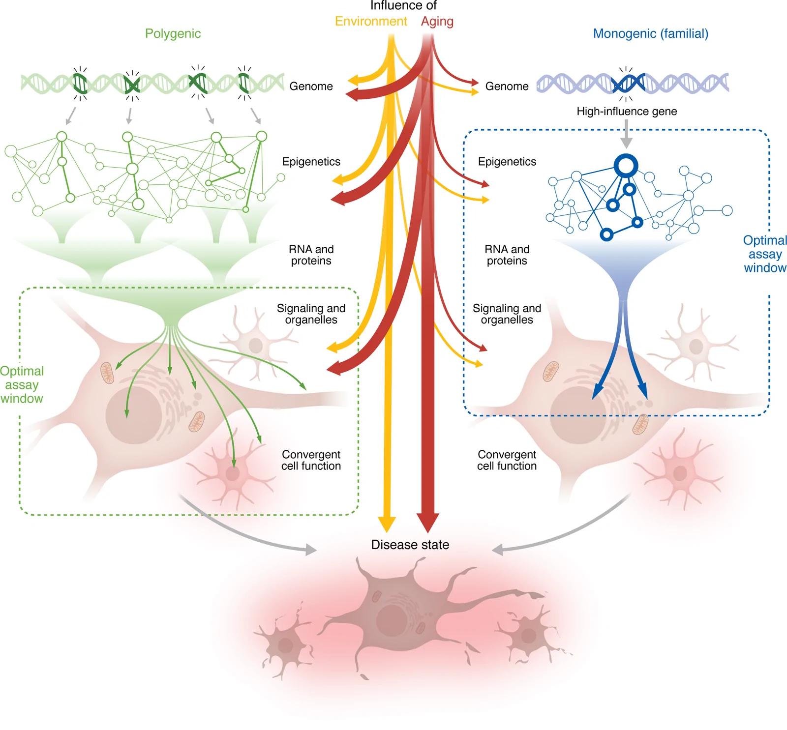
A review in the Journal of Clinical Investigation examines how human induced pluripotent stem cell (iPSC) systems can support neurodegeneration research and therapeutic discovery. Carrying a disease-associated genotype does not guarantee a measurable cellular phenotype. Patient-derived iPSCs preserve the donor’s wider genetic background, which is useful when risk comes from many variants rather than one dominant mutation. Engineering a defined mutation into a common reference line reduces background variation, but it answers a different question. A phenotype in that controlled background need not capture the range of responses among patients who carry the same variant.
The authors link this distinction to assay choice. A high-penetrance familial mutation may produce its clearest signal near the initial molecular defect, before downstream compensatory responses obscure it. For polygenic disease, individual effects may be too small to detect reliably on their own. A downstream cellular process at which several effects converge may provide a more measurable endpoint. The appropriate readout therefore depends on the architecture of the disease as well as on the available culture technology.
An observable change also needs a biological interpretation. The review distinguishes pathogenic processes from incidental changes and protective adaptations. Reducing a conspicuous cellular feature would not necessarily be beneficial if that feature were part of the cell’s response to stress. For therapeutic screens, this makes the relationship between the measured phenotype and the disease process as important as how reproducibly a drug changes the readout.
Choose the Culture for the Question
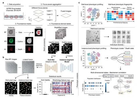
Two-dimensional cultures of a defined neuronal subtype make cell-intrinsic effects easier to measure and are more compatible with larger screens. Adding astrocytes, microglia or oligodendrocytes lets researchers examine interactions that a single-cell-type culture cannot represent. Three-dimensional organoids add spatial organization and some features of tissue maturation, but they take longer to grow, vary between batches and are less straightforward to quantify at scale.
The authors argue for choosing the simplest model that contains the biology needed for a particular question. Greater complexity is useful when the question depends on interactions or tissue architecture; it is not an automatic improvement for every drug screen. Organoids may reach more mature states than two-dimensional cultures for some features, yet the review notes that even long-lived organoids generally remain closer to fetal or early postnatal than adult brain tissue.
Aging Does Not Return Just by Adding Complexity
Reprogramming a donor cell to the pluripotent state resets much of its epigenetic age. That feature is useful for expanding patient-derived cells but can erase the cellular history relevant to late-onset disease. Culture complexity and biological age therefore require separate consideration.
The aging approaches have different interpretations. Progerin expression, telomere manipulation and chemical induction of senescence can expose vulnerabilities that are muted in young iPSC-derived neurons. Environmental stressors can likewise test how a genetic background responds to inflammation or metabolic challenge. The review treats these as ways to model selected pressures or sensitize a system, because each can bias the experiment toward the pathway being perturbed. A response to one induced stress is not equivalent to the full cellular history of an older donor.
Direct conversion turns donor somatic cells into neurons without a pluripotent intermediate, preserving age-associated methylation patterns, DNA damage and other donor-cell features. That advantage comes with a practical constraint: primary somatic cells have limited expansion, and repeatedly expanding or resetting them can interfere with the aged state the model is intended to retain. The low yield also constrains large screens and gene-corrected isogenic controls. Donor variation becomes part of the design, requiring enough independently sampled people to distinguish disease-associated differences from variation in biological age. The review presents this as a distinct experimental choice, especially for late-onset phenotypes, rather than a universally superior replacement for iPSC models.
Implications for Drug Research
A neuron-intrinsic screen, a test of glial contributions and a study of late-onset pathology may require different human-cell systems. The review provides a framework for making those choices, rather than a direct comparison showing that any one model predicts clinical drug response better. The relevant cellular property—genetic background, interaction with other cell types, tissue organization or donor age—needs to be present for the experiment to address the intended therapeutic question.