Reconstituting Tumor, Stroma and Immune Cells
A Seoul National University-led team built renal cell carcinoma (RCC) assembloids from separately expanded tumor cells, cancer-associated fibroblasts (CAFs) and tumor-infiltrating immune cells. The autologous components were combined at proportions reflecting each parental tumor. This design allowed individual cell populations to be exchanged, removed or genetically altered.
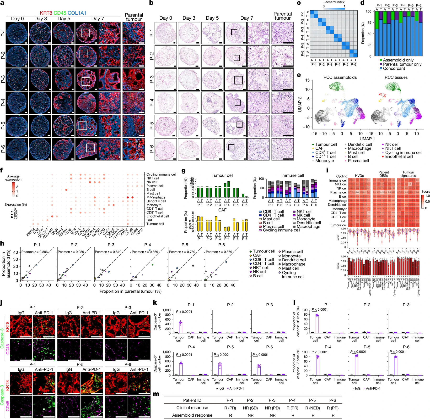
The initial models came from six pretreatment patient specimens. Multicellular organization emerged by day 5 and reproduced selected parental histological and cellular features by day 7, with viability above 90% during assembly. Genomic and single-cell comparisons examined the tumor alterations and microenvironmental compartments retained in culture.
After assembly, a further seven days of 10 µg/mL anti-PD-1 or matched isotype control produced tumor-cell reduction and increased apoptosis in four patient models, while two showed no significant tumor-cell death. Apoptosis was concentrated in tumor cells, with comparatively little in CAFs and immune cells. Responses mirrored the corresponding patients’ immune-checkpoint outcomes within this small cohort.
Component Exchange Identifies a Supportive Niche
Responsive models contained more stem-like CD8+ T cells marked by TCF1 positivity and TIM3 negativity. Mixing responder and nonresponder components, and removing individual populations, showed that maintenance of this state depended on tumor cells and CAFs together. The immune cells’ origin alone did not determine the response.
Lineage tracing attributed maintenance of the stem-like population mainly to expansion of pre-existing cells. Conditioned-medium experiments placed CAFs between tumor-derived soluble signals and T-cell support: tumor-conditioned medium restored the relevant state in tumor-deficient assembloids that still contained CAFs, whereas CAF-conditioned medium alone did not replace missing CAFs. The component-based design allowed this sequence of interactions to be tested.
Candidate-factor tests linked tumor-derived WNT2B to CAF-derived activin A. Knocking down INHBA in CAFs depleted stem-like T cells and abolished anti-PD-1 responsiveness, whereas knockdown of the also-upregulated FGF2 did not have the same effect. Tumor-cell WNT2B knockdown reduced CAF INHBA expression, stem-like T cells and antibody response. These perturbations connected the inferred signaling relationship to function.
Resistance Reflects the Stromal Response to WNT
Tumor WNT2B expression was similar in responders and nonresponders. Nonresponder CAFs instead had lower LRP6, impaired WNT signaling and reduced activin A expression. Responder tumor cells could not restore this behavior when paired with the deficient CAFs. Reintroducing LRP6 into nonresponder CAFs restored INHBA expression, stem-like CD8+ T cells and anti-PD-1 sensitivity.
Additional air–liquid-interface organoids supported the axis: WNT inhibition removed antibody responsiveness in a responder-derived model, while recombinant activin A restored sensitivity in a nonresponder-derived model. Separate humanized-mouse experiments provided further mechanistic evidence.
An independent eight-sample pretreatment single-cell cohort and additional spatial analyses linked LRP6/INHBA-associated CAF features to T-cell stemness and treatment response. Responsive tumors more often placed these stromal features near tumor cells and the relevant T-cell populations. These tissue observations provide a reference for the interactions tested in the assembloids. Overall CD8+ T-cell infiltration was comparable in the parental tissues, while responder tumors were enriched for stem-like T cells and local LRP6-positive CAFs.
A Mechanistic Platform With Defined Limits
The platform can test how individual tumor, stromal and immune components affect a treatment response, rather than measuring aggregate survival alone. Its strength is the combination of patient-derived material, controlled component exchange and functional perturbation.
Culture selection remains a limitation. The minor CA9-negative compartment may include non-neoplastic epithelial cells and may change during expansion. Other microenvironmental populations were not comprehensively tested. The proposed role of activin A also depends on context: differences between inflamed and immune-cold tumors, and the timing of pathway modulation, were not directly established here.
Concordance in six patients and the mechanistic experiments support further use of the system for research. They do not establish clinical predictive performance or benefit from manipulating this pathway in patients. Related patterns in public datasets from other cancers remain associative and require validation for each intended setting.