Evaluate checkpoint-inhibitor responses and identify contributions from stromal behavior and T-cell state.
Model & Design
Pretreatment RCC CA9-positive/negative cells, autologous CAFs and tumor-infiltrating immune cells, expanded separately and reconstituted at parental-tumor proportions.
Experimental Format
Seven-day assembly followed by seven days of 10 µg/mL anti-PD-1 or matched IgG1. This excludes earlier isolation and expansion.
Model Components
Component
Autologous cellular components
Description
Patient-derived tumor cells, CAFs and infiltrating immune cells; CA9-negative cultures may contain non-neoplastic cells.
Component
Response and mechanism comparisons
Description
Isotype control, component exchange/removal, WNT2B or INHBA knockdown and CAF LRP6 restoration; flow cytometry and cell-specific histological imaging.
Measurements & Endpoints
Tumor-cell reduction and cleaved-caspase-3-positive apoptosis
TCF1+TIM3− CD8+ T-cell proportion
CAF INHBA/LRP6 and WNT responsiveness
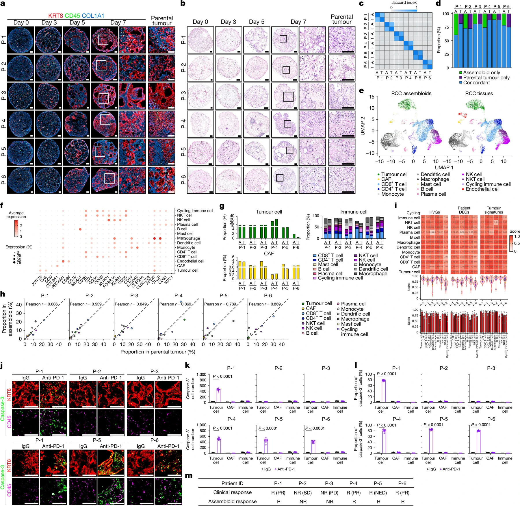
Concordance with parental composition and patient treatment response
Analysis
Preserve patient, independent-assembloid and section units. Interpret autologous, exchanged-component and genetic-perturbation conditions separately using tumor and immune endpoints.
Published Evidence
Study Design
Autologous antibody-response comparison with component and signaling perturbations.
Study Sample
Six patients. Main imaging used three independent assembloids per condition and three sections each.
Comparison
Anti-PD-1 versus IgG1; autologous or exchanged components; corresponding genetic controls.
Finding
Four patient-derived models responded and two showed no significant tumor-cell death, matching patient-response directions. WNT2B/INHBA perturbation impaired responses, while LRP6 restoration recovered responses with nonresponder CAFs.
Interpretation & Limits
Six-patient concordance does not establish prospective clinical predictive performance.
Culture selection, CA9-negative-cell identity and other microenvironmental contributions remain unresolved.
Activin A effects across tumor/inflammatory settings and intervention timing require separate validation.
Assembloids from six patients reproduced their corresponding immunotherapy-response patterns. Component exchange and perturbations connected tumor WNT2B, fibroblast activin A and stem-like CD8 T cells, with low stromal LRP6 linked to resistance.