
Single-Cell Colorectal Organoid Arrays Separate Growth Inhibition from Cell Killing
A four-patient microfluidic study measured drug responses in individual single-cell-derived organoids. Transcriptome-selected compounds often slowed growth without strong killing; selected combinations improved clearance in the assay.
Keeping Individual Tumor-Cell Descendants Separate
Researchers at South China University of Technology, Guangzhou First People’s Hospital and collaborating academic centers developed a microfluidic array for single-cell-derived colorectal tumor organoids. Their Microsystems & Nanoengineering study used the separate organoid units to expose response differences that could be obscured when multiple starting cells contribute to one culture.
The chip contained 3,618 microwells. Stochastic capture produced approximately 1,000 single-cell culture positions under the chosen loading condition. An alginate–chitosan membrane retained the cells while allowing molecular exchange, and 20% Matrigel supported organoid formation. The resulting structures were followed by imaging rather than pooled into a single average.
The Model Preserved Tumor Features While Selecting Surviving Cells
The experiments used tumor material from four patients. Single-cell organoid formation rates varied among these sources, from approximately 19% to 36%. Most starting cells therefore did not form an organoid. The method preferentially expanded cells able to survive and self-renew in the culture environment.
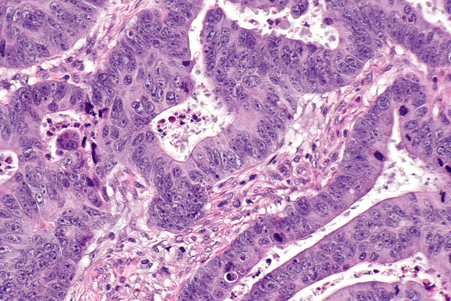
Histological comparisons identified solid and luminal structures, with CK20, CDX2, MUC2 and chromogranin A staining supporting differentiated colorectal epithelial features. Whole-exome comparisons found more than 85% concordance of driver mutations between the single-cell-derived organoids and corresponding tumor tissue. This characterization supports the models’ tumor origin while leaving the selective nature of establishment relevant to interpretation.
The investigators also made multicell-derived organoids as a comparator. Single-cell-derived organoids grew for seven days before drug testing, whereas the multicell-derived models grew for three days. Differences between them consequently reflect both starting-cell organization and the particular assay schedules.
A Chemotherapy Challenge Revealed Resistant Subpopulations
The study modeled a FOLFIRINOX-related cytotoxic combination using 40 μM 5-fluorouracil, 125 nM SN38 and 5 μM oxaliplatin. SN38 replaced the prodrug irinotecan. Treatment lasted three days under static conditions, with fresh drug solution supplied daily. Calcein-AM and propidium iodide images supplied live/dead signals, while automated analysis also measured organoid size.
Individual single-cell-derived organoids showed a wider viability distribution after treatment, reported as 0–70%, compared with 65–100% in multicell-derived organoids. Smaller single-cell-derived structures were relatively resistant compared with larger ones. These distributions matter because a reduction in average size can coexist with surviving individual units.
Resistant organoids were recovered for transcriptomic analysis. Pathway and prognosis-associated gene analyses identified patient-dependent features, often linked to PI3K–AKT and related signaling. The oncoPredict R package, trained on existing cell-line expression and drug-response resources, then nominated candidate compounds. These computational predictions were followed by direct tests on the organoid arrays.
Candidate Combinations Improved Clearance in the Array
The initial screen tested 34 candidate compounds at 5 μM. All showed antiproliferative effects by size measurement, but only a subset showed significant cytotoxic effects by viability. When retested on resistant organoids, the compounds continued to suppress growth while their killing effects were limited. Growth inhibition and elimination of the resistant units therefore remained separate outcomes.
Adding selected candidates to the chemotherapy combination reduced both size and viability more strongly than chemotherapy alone. Separate concentration-response tests identified lower IC50 values with combination treatment. A final clearance comparison used selected compounds at 50 μM for three days and found fewer organoids retaining viability above 50%. The patient-specific candidates were IMD-0354, AT7519, teniposide and GNE-317 for the four respective models. Their combination indices were below 1 under the study’s analysis.
The screening and dose-response experiments were repeated twice, and no prospective comparison with patient treatment outcomes was reported. The work establishes a way to couple within-culture response distributions to molecular hypotheses and follow-up drug tests. Whether the nominated combinations improve treatment in patients remains outside the evidence from this four-patient array study.