
Electrode-Anchored Tumor Spheroids Reveal Early Drug-Response Signals in Impedance
A scaffold-integrated electrode array tracked maturation and drug responses in U-87 MG spheroids. Early impedance changes preceded significant staining changes after nutlin-3a, while older cultures and additional drug tests showed why electrical signals require biological and exposure context.
Anchoring a Three-Dimensional Culture to Its Sensor
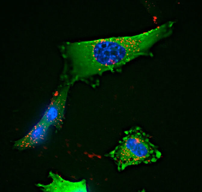
Researchers at the Italian Institute of Technology and academic collaborators developed an electrode array that keeps tumor spheroids at defined sensing sites. The study used U-87 MG glioblastoma cells expressing green fluorescent protein (GFP), with a separate CaCo-2 colorectal-cancer application. The platform combines three-dimensional culture and repeated electrical measurements without requiring a microfluidic flow circuit.
Cupola-shaped microscaffolds fabricated by two-photon polymerization guide cells onto the measurement electrodes. A different tower-shaped structure protects the reference electrode from colonization. In the planar comparison, migrating cell clusters rarely stayed on the sensing surface. At 48 hours, the fraction of responsive electrodes was 13 ± 4% in two-dimensional cultures, compared with 81 ± 10% with three-dimensional scaffolds; the reported groups comprised 16 and 26 electrodes, respectively.
The 16-electrode array supports multiplex measurement, while optical access permits complementary microscopy. The scaffold changes the contact between cells and sensor, so improved electrical coverage is a distinct result from establishing that every impedance change represents cell death.
Culture Age Changes the Nutlin-3a Response
The investigators compared spheroids matured for 48 or 96 hours before treatment with 30 μM nutlin-3a, an antagonist of the p53–MDM2 interaction. Measurements after 3 and 24 hours showed reduced impedance with either an external reference electrode or the protected integrated reference. Younger cultures approached cell-free electrode values, whereas older cultures retained more signal and showed a progressive response over time.
Additional testing in 96-hour spheroids separated dose effects. At 10 or 15 μM, nutlin-3a prevented further impedance growth but did not produce a significant decrease by 24 hours. At 20 μM, impedance fell significantly. These observations distinguish a suppressed growth-associated signal from the larger disruption produced at higher exposure.
Biological assays supplied an important timing comparison. After 3 hours of 30 μM nutlin-3a, impedance had fallen, but neither the proportion of membrane-compromised EthD-1-positive cells nor p53-positive cells increased significantly. At 24 hours, the respective proportions reached 48.6 ± 6.7% and 35.6 ± 6.8%. Detachment could contribute to an electrical change while removing cells from the population available for staining; the early signal therefore has more than one possible biological contribution.
For the early staining comparison, EthD-1 positivity was 16.6 ± 4.4% after treatment versus 13.8 ± 4.9% in controls; p53 positivity was 17.7 ± 4.0% versus 14.6 ± 2.0%. The paper reports these measurements as mean ± standard deviation. Their nonsignificant early differences constrain the biological interpretation of the electrical change.
Frequency and Drug Exposure Define the Comparison
Untreated spheroids also changed electrically with age. In a 20-spheroid analysis, seven-day cultures showed a low-frequency decline around 10–100 Hz alongside central EthD-1 staining. Removing cells returned impedance toward cell-free values, supporting a cellular rather than electrode-failure origin. The authors associate this pattern with loss of core viability and changing tissue structure.
Temozolomide produced smaller changes: the device experiment, reported at a nominal 30 mM, reduced cell index by about 5.7% at 24 hours and 11.5% at 48 hours. A separate non-adherent spheroid flow-cytometry experiment used 30 μM for 48 hours and reported a 13.3% increase in annexin-V-positive cells. Because those exposures differ, the two assays provide separate observations rather than a matched-dose calibration.
In the CaCo-2 extension, 96-hour spheroids received 15 μM sorafenib. Cell index fell by approximately 37% after 24 hours, demonstrating a response in a second cell-line tumor model.
A Repeated Measurement with an Aggregate Biological Meaning
The study supports repeated, label-free monitoring of sensor-anchored tumor cultures, with exposure, maturation and frequency all affecting interpretation. Impedance integrates adhesion, cohesion, membrane properties and cell loss across the measured structure. It does not resolve individual cell states, and the study did not use equivalent-circuit modeling to separate those contributions.
Patient-derived models, stromal or immune cocultures and softer scaffold materials were proposed for further development. The present evidence comes from established cell lines and defined drug experiments; its immediate drug-evaluation value is the ability to detect a changing electrical phenotype and then connect that phenotype to appropriately timed biological measurements.