BBB Chip Drug-Exposure Monitoring Coupled to Norepinephrine Secretion
Human cell-line barrier–neural cultures pair pramipexole concentration measurements with evoked norepinephrine after chemical injury.
Brain & Neural ModelsOrgan Chips
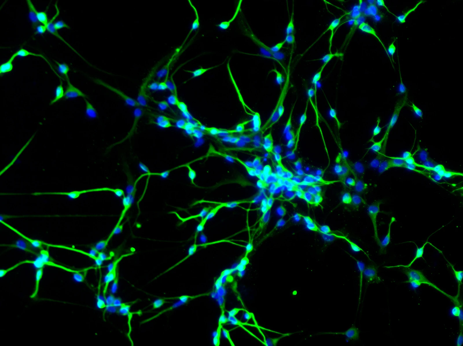
Fluorescence micrograph of neurons differentiated from human neural stem cells. (Yirui Sun, CC BY 4.0)
Research Applications
Relate trans-barrier drug arrival to a functional secretory response and compare microfluidic with static transfer.
Model & Design
hCMEC/D3 human endothelial cells and undifferentiated SH-SY5Y human neuroblastoma cells on opposite collagen-I/fibronectin-coated PET faces; no astrocytes/pericytes.
Experimental Format
120 μL/min dynamic culture; 24-hour 3 mM 1-methyl-4-phenylpyridinium (MPP+) injury then 24-hour pramipexole exposure; static Transwell comparator.
Model Components
Component
Cells and exposure
Description
7.5×10^5 endothelial and 1×10^5 SH-SY5Y cells. Vascular pramipexole 750 μM; selected cell-response benchmark 200 μM. Injury-free, injured/no-drug and nutrient-replenishment controls.
Component
Measurement system
Description
Electrochemical concentration sensor with regeneration and LC–MS drug comparison; separate LC–MS evoked norepinephrine assay. The drug sensor does not measure norepinephrine online.
Measurements & Endpoints
Neural-side pramipexole concentration and permeability
Evoked norepinephrine secretion
CCK-8 metabolic activity and apoptosis
ZO-1 and 4/40-kDa dextran permeability
TNFα, IL-1β and IL-6
Analysis
Norepinephrine secretion and CCK-8 metabolic activity are separate response measures. Reported ± values are mean ± standard deviation (SD). Total clock times of 37 and 32 hours include the preceding 24-hour injury period, corresponding to approximately 13 and 8 hours after drug dosing.
Published Evidence
Study Design
Dynamic versus static barrier transfer, with orthogonal drug concentration detection and chemical-injury response comparisons.
Study Sample
At least three independent experiments in source statistics; barrier Figure 5 n = 6–8 by condition; no donor cohort.
Comparison
Chip versus Transwell, injured/no-drug versus pramipexole, normal versus injured cells and nutrient controls.
Finding
Pramipexole transfer was slower in chip than Transwell (Papp 7.28×10^-7 versus 1.22×10^-6 cm/s). Norepinephrine improved after dosing; apoptosis 19.95 ± 1.06% fell to 13.95 ± 1.63%. TNFα declined but IL-1β/IL-6 were unchanged; 6 mM MPP+ injury resisted rescue. 40-kDa permeability did not increase significantly with the selected injury.
Interpretation & Limits
Undifferentiated neuroblastoma cells are not normal noradrenergic or dopaminergic neurons; evoked product was predominantly norepinephrine.
High experimental pramipexole concentrations and acute MPP+ injury are not clinical exposure predictions.
Drug sensing is in situ but functional secretion requires offline LC–MS sampling.
Sensor fouling reduced the fifth uncleaned signal to 47.6%; regeneration was necessary for repeat measurements.
A human cell-line barrier–neural chip coupled drug concentration measurements with norepinephrine secretion after MPP+ injury. Pramipexole crossed more slowly than in static Transwells, and functional recovery differed from metabolic viability and inflammatory readouts.