
A Blood–Brain Barrier Chip Tracks Pramipexole Exposure and Norepinephrine Secretion
A human cell-line barrier–neural chip coupled drug concentration measurements with norepinephrine secretion after MPP+ injury. Pramipexole crossed more slowly than in static Transwells, and functional recovery differed from metabolic viability and inflammatory readouts.
Drug Concentration and Cellular Function Were Measured Separately
A Fujian Medical University study in Microsystems & Nanoengineering coupled a blood–brain barrier chip to an electrochemical drug sensor and liquid chromatography–mass spectrometry, or LC–MS. The investigators used pramipexole to examine how transport across an endothelial layer relates to recovery of neurotransmitter secretion after a chemical injury. Measuring concentration and function separately allowed the experiment to distinguish drug arrival from the timing of a cellular response.
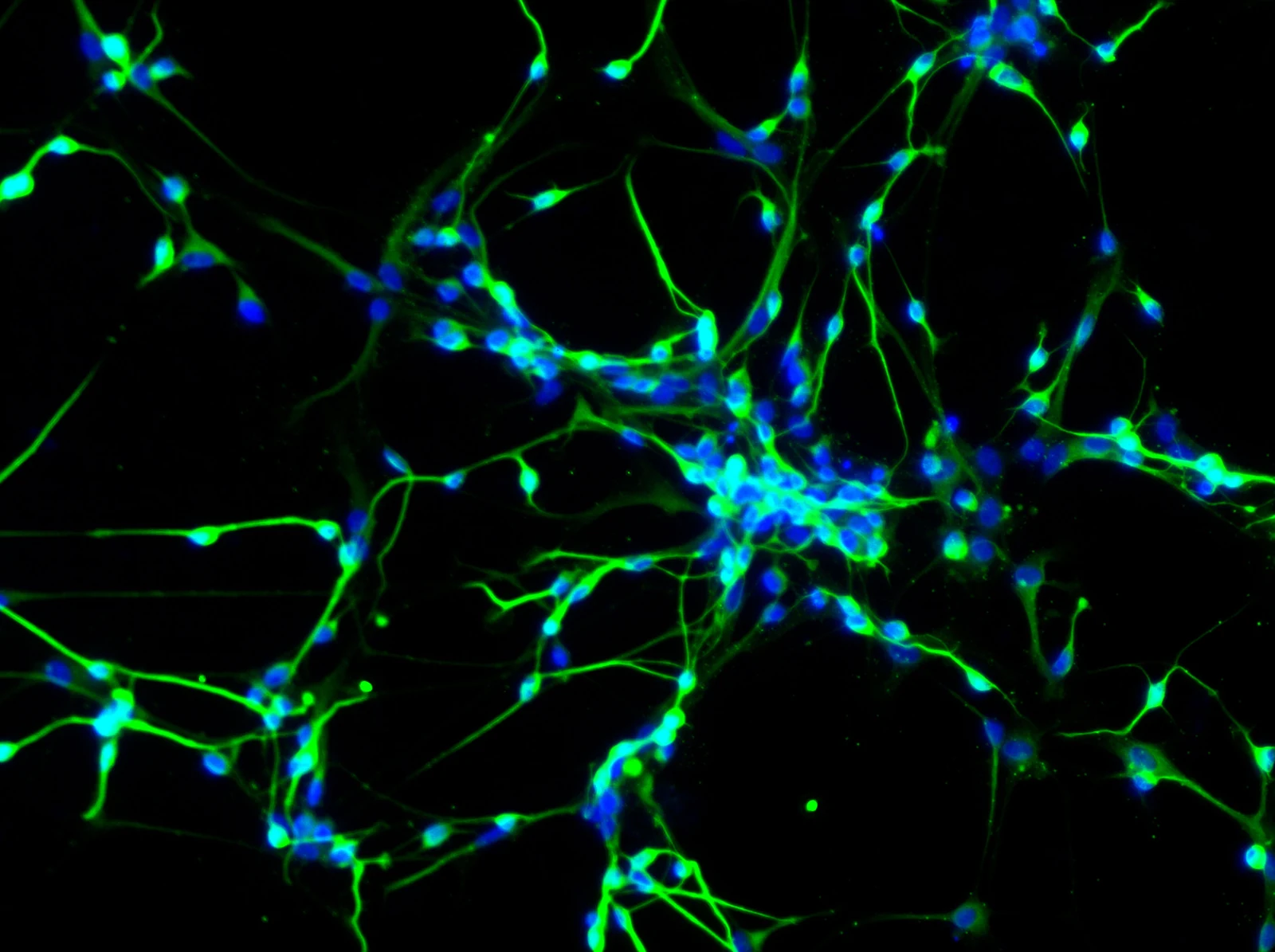
The barrier consisted of the human endothelial cell line hCMEC/D3, while the neural compartment used undifferentiated SH-SY5Y human neuroblastoma cells. Cells occupied opposite faces of a collagen-I/fibronectin-coated porous membrane. The circuit ran at 120 μL/min. This was a two-cell-line model, without astrocytes, pericytes or normal patient-derived neurons.
Dynamic culture improved junctional staining and reduced 40-kDa dextran permeability relative to static conditions. Electrical resistance reached 134.0 ± 22.4 Ω·cm²; reported uncertainty values in this study are mean ± standard deviation (SD). Transporter-inhibitor experiments supported P-glycoprotein and multidrug resistance-associated protein activity, and hypertonic mannitol reversibly opened the barrier. These measured features supplied a barrier context for the drug-transfer experiment.
The Injury Readout Was Primarily Norepinephrine
The team compared retinoic-acid-differentiated and undifferentiated SH-SY5Y cells, then selected the undifferentiated cells for the chip. The neurotoxic compound 1-methyl-4-phenylpyridinium (MPP+) was applied for 24 hours. Evoked secretion consisted predominantly of norepinephrine, with little dopamine or epinephrine detected. Norepinephrine therefore served as the functional readout.
The concentration associated with a half-maximal loss of the norepinephrine response was 3.71 ± 0.36 mM MPP+, compared with 6.72 ± 0.43 mM for the CCK-8 metabolic assay. The difference shows that secretory impairment emerged at an exposure where metabolic activity gave a less severe injury estimate. The researchers selected 3 mM for subsequent modeling; 6 mM caused extensive damage and could not be rescued by the tested pramipexole conditions.
Pramipexole at 200 μM improved metabolic and secretory responses after the lower injury exposure. In a separate post-injury comparison, apoptosis fell from 19.95 ± 1.06% to 13.95 ± 1.63%. TNFα declined, while IL-1β and IL-6 did not change significantly. Thus, the measured response was neither complete recovery nor uniform suppression of inflammatory markers.
The Chip Delayed Drug Transfer Relative to a Transwell
For exposure–response monitoring, the vascular compartment received 750 μM pramipexole after 24 hours of injury, followed by another 24 hours of observation. Electrochemical measurements and LC–MS tracked the drug on the neural side; LC–MS separately quantified evoked norepinephrine secretion. The electrochemical sensor measured drug concentration in situ, while the functional secretory assay required sample collection.
The neural-side pramipexole concentration reached the selected 200 μM response benchmark about 13 hours after dosing in the chip, compared with about eight hours in a static Transwell. The source reports these as total experimental clock times of approximately 37 and 32 hours, including the preceding 24-hour injury period. Apparent permeability was 7.28 × 10⁻⁷ ± 3.53 × 10⁻⁷ cm/s in the chip and 1.22 × 10⁻⁶ ± 3.53 × 10⁻⁷ cm/s in the Transwell.
Norepinephrine secretion continued to improve after that concentration benchmark was reached. Serum-containing medium alone also improved responses in uninjured controls, making nutrient replenishment a relevant comparator. After MPP+ injury, pramipexole increased ZO-1 staining and reduced 4-kDa dextran permeability; 40-kDa permeability had not significantly increased with injury. The assays support size-dependent barrier changes rather than an undifferentiated claim of barrier breakdown and repair.
Repeated Sensing Required Regeneration
The integrated sensor was linear over 5–500 μM pramipexole. Repeated measurements without cleaning lost signal: by the fifth measurement, the response was 47.6% of its initial value. Electrochemical regeneration reduced the relative standard deviation across five measurements to 4.46%, with a similar 4.84% value in the chip. This correction was essential to longitudinal concentration monitoring.
The platform connects drug transfer with a functional secretory endpoint in a controlled human cell-line system. Its interpretation remains tied to high experimental pramipexole concentrations, acute MPP+ injury and a simplified barrier. The neuroblastoma origin and absence of supporting neurovascular cells are especially relevant when extending the measured norepinephrine response to disease-specific human neurons.