Colorectal Cell-Line Organoid Washout and Regrowth Assay
Follow residual PLR123 and LS174T three-dimensional cultures through SN-38 exposure, washout and regrowth, then test pathway-directed inhibitors.
Tumor ModelsOrganoids
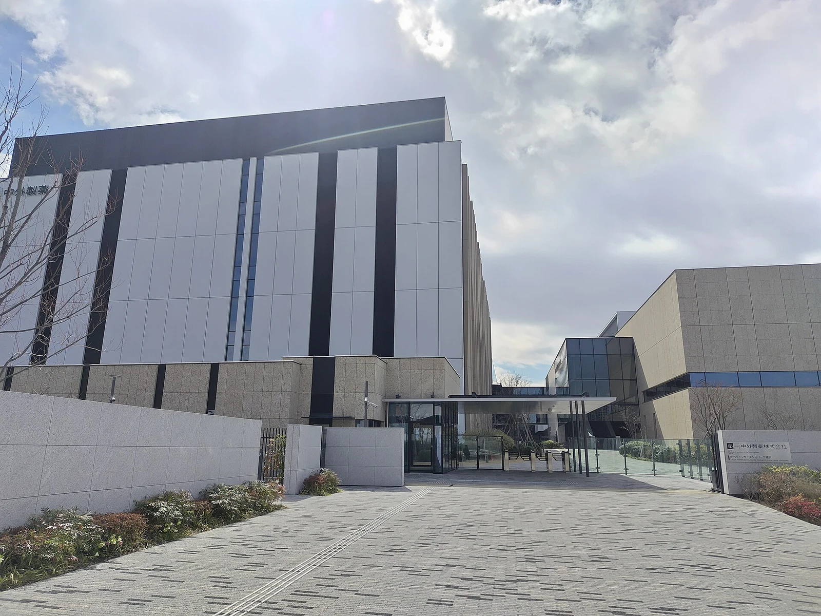
Chugai Life Science Park Yokohama, photographed in March 2026. (妖精書士, CC0 1.0)
Research Applications
Identify dependencies of chemotherapy-tolerant residual cells and distinguish survival from the transition back to proliferation.
Model & Design
PDX-derived PLR123 colorectal-cancer cells with stem-cell properties and separate LS174T cell-line-derived organoids in Matrigel. Twelve archived clinical specimens provide a tissue comparison.
Experimental Format
SN-38 at 300 nM from culture day 10 to 13, followed by washout and re-embedding in fresh Matrigel. Time-course morphology and transcriptomics are paired with inhibitor-specific regrowth endpoints.
Model Components
Component
Cell-state measurements
Description
Ki-67, LGR5, HMGCS2 and p21 staining; bulk and single-cell RNA sequencing before treatment, during treatment and on regrowth days 2, 4 and 6.
Component
Interventions
Description
XAV-939 10 μM; BSO 200 μM and auranofin 200 nM alone/together; separate harmine, mitochondrial-inhibitor and BMH-21 comparisons. The LS174T comparison in Figure 7I specifies BMH-21 at 3 μM.
Measurements & Endpoints
Residual-culture viability
Regrowth morphology and proliferation
LGR5-positive fraction
Cell-state expression and inferred trajectories
Analysis
Separate changes in marker expression from dependence established by perturbation. Match the sample, cell line, inhibitor dose and readout day for each experiment; mouse lung-nodule findings are separate corroboration.
Published Evidence
Study Sample
Two established colorectal-cancer lines; 12 clinical specimens for morphology and markers. Regrowth-day-2 single-cell analysis includes 1,698 cells; individual culture assays have separately reported repeat counts.
Comparison
Pretreatment, drug and washout time points; vehicle versus pathway inhibitors; single versus combined redox inhibition; separate irinotecan-treated mouse lung-nodule models.
Finding
Regrown PLR123 retained SN-38 sensitivity. XAV-939 limited regrowth; BSO/auranofin were inactive individually but active together. HMGCS2 knockdown and mitochondrial inhibitors did not change the tested phenotype. BMH-21 eliminated residual cells in culture and limited regrowth in mice; XAV-939 and harmine culture hits did not produce significant added mouse growth inhibition.
Interpretation & Limits
Two cell-line-derived systems and selected archived tissue comparisons.
RNA velocity infers transitions rather than tracing lineages.
Inhibitor conditions and endpoint days differ; no single common exposure regimen.
Mouse burden reduction does not establish clinical recurrence prevention; combination dosing caused reversible body-weight loss.
Study Design
Primary study comparisons of the named models, exposures and measured outcomes.
Cell-line-derived organoids resumed growth after transient SN-38 exposure and reproduced selected features of residual colorectal tumors. Time-resolved profiling identified distinct non-cycling states; RNA polymerase I inhibition suppressed regrowth in culture and mouse experiments, while some other culture hits failed in vivo.