
A Colorectal Cancer Regrowth Model Separates Residual-Cell States and Drug Vulnerabilities
Cell-line-derived organoids resumed growth after transient SN-38 exposure and reproduced selected features of residual colorectal tumors. Time-resolved profiling identified distinct non-cycling states; RNA polymerase I inhibition suppressed regrowth in culture and mouse experiments, while some other culture hits failed in vivo.
Modeling What Survives Chemotherapy
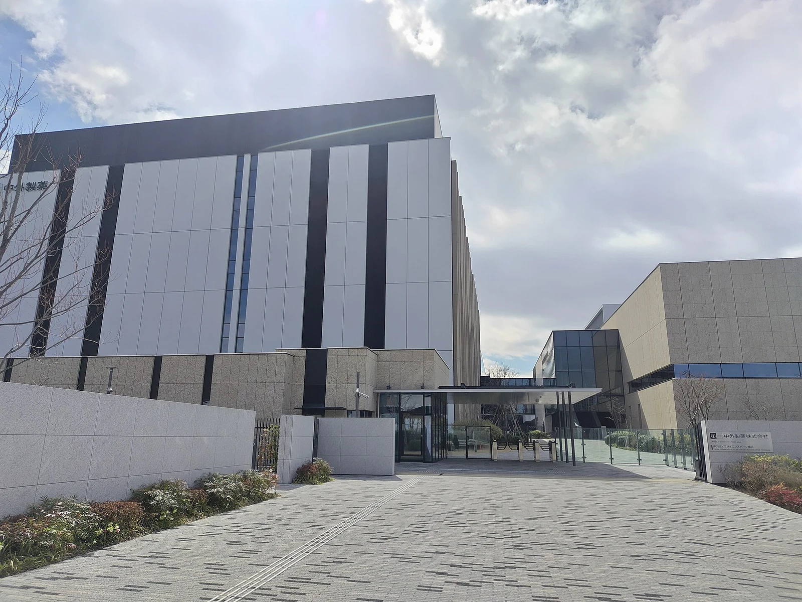
A Chugai Pharmaceutical-led study with academic collaborators developed a three-dimensional model of colorectal cancer regrowth after chemotherapy. Its starting clinical observation was a series of 12 archived surgical specimens collected after neoadjuvant treatment. Small epithelial clusters remained within fibrous tissue and were largely negative for the proliferation marker Ki-67, despite expression of stem-cell-associated markers.
The residual clinical clusters retained epithelial E-cadherin and lacked the tested vimentin and ZEB1 signals. LGR5 staining was high in four specimens and detectable in seven of the remaining eight, so the clinical reference itself contained heterogeneous marker states.
The experimental cultures were derived from established cells rather than newly generated organoids from those 12 patients. PLR123 is an LGR5-positive colorectal-cancer line originating from a patient-derived xenograft. LS174T provided a separate commercially available cell-line comparison. Both formed structures in Matrigel that allowed residual-cell survival and renewed proliferation to be examined over time.
In the main model, cultures received 300 nM SN-38, the active metabolite of irinotecan, for three days. Surviving aggregates were washed and re-embedded in fresh Matrigel without drug. This produced more reproducible regrowth timing than a medium change alone.
Non-Cycling Cells Occupy Several States
Residual aggregates initially contained many Ki-67-negative cells. By regrowth day 2, small clusters included proliferating and LGR5-positive cells, and by day 6 proliferative structures had expanded. LGR5-positive fractions changed from 4.1% before treatment to 14.7% during treatment, 58.5% on regrowth day 2 and 37.5% on day 6. Regrown PLR123 cells retained sensitivity to SN-38, consistent with a reversible tolerant state rather than demonstrated stable genetic resistance.
Bulk and single-cell RNA sequencing followed pretreatment, drug exposure and several regrowth time points. The analysis separated cycling populations from non-cycling populations with DNA-damage, metabolic or translation-associated programs. HMGCS2, a ketone-synthesis enzyme, marked one state; its expression was also found in residual clusters in 7 of the 12 clinical specimens, with substantial variation between clusters.
A closer analysis of 1,698 cells on regrowth day 2 identified a non-cycling group with high expression of translation-related genes including RPL17 and EEF1G. RNA-velocity analysis positioned this group upstream of other regrowth-associated states. This supplies a hypothesis about state transitions from transcript dynamics; it does not directly trace the fate of individual cells.
Culture Screening Distinguishes Regrowth Suppression from Cell Elimination
The researchers tested pathways highlighted by the profiles. The Wnt-pathway inhibitor XAV-939 reduced regrowth and proliferation-associated gene expression, but residual cells remained. Depleting glutathione with buthionine sulfoximine (BSO) or inhibiting the thioredoxin system with auranofin had little effect individually, whereas the combination was active. Harmine also reduced residual-cell survival.
Other apparently relevant molecular changes did not yield equivalent vulnerabilities. HMGCS2 knockdown did not change regrowth, and the mitochondrial respiration inhibitors oligomycin and FCCP did not induce cell death under the tested conditions. By contrast, the RNA polymerase I inhibitor BMH-21 eliminated residual cells in the culture experiments. The LS174T model reproduced several response patterns and the strong BMH-21 result.
The Mouse Comparison Narrows the Translational Claim
A separate model used intravenously implanted PLR123 cells to generate lung nodules in immunodeficient mice. Irinotecan reduced proliferation, followed by renewed expansion during a drug-free interval. Sequential or cyclic inhibitor experiments then tested whether the culture-derived vulnerabilities persisted in this setting.
XAV-939 and harmine did not significantly reduce tumor size beyond irinotecan alone, despite their culture activity. BMH-21 reduced tumor burden with either schedule and reduced subsequent nodule size and number. Lower Ki-67 staining, with similar cleaved-caspase-3 staining, supported suppression of the return to proliferation in the sampled tumors. Combination treatment caused about 10% body-weight loss during dosing, followed by recovery during the drug-free period.
The work offers a controlled way to investigate early regrowth and to reject targets whose expression changes do not translate into functional dependence. Its clinical-tissue comparison supports selected morphological and marker similarities, while drug efficacy remains evidence from two cell-line systems and mouse models. It does not establish elimination of residual disease or prevention of recurrence in patients.