Mucinous Gastric Cancer Organoid Mucin and Drug-Response Measurements
Combine subtype-specific mucin imaging and tumor characterization with a chemotherapy concentration-response panel.
Tumor ModelsOrganoids
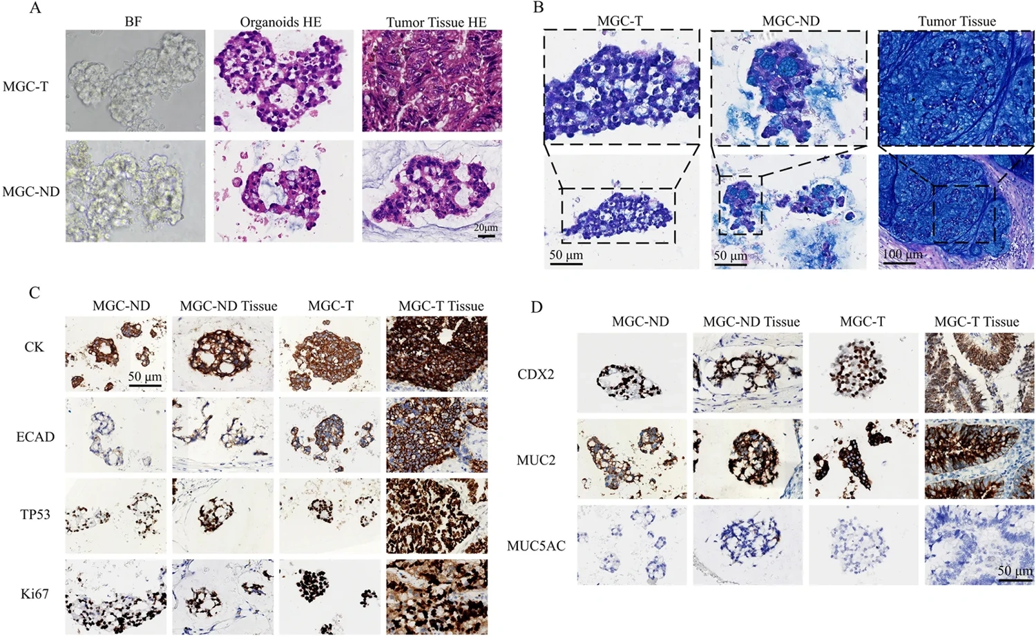
Matched mucinous gastric tumors and organoids show morphology, mucin staining and tumor-marker expression. (Haobin Hou et al., CC BY 4.0)
Research Applications
Investigate pharmacological differences among mucinous gastric tumors and between matched primary and metastatic tissue.
Model & Design
Four mucinous gastric cancer organoid models from three patients, including one matched primary–peritoneal-metastasis pair.
Experimental Format
72-hour CellTiter-Glo 2.0 ATP-associated viability across an 11-agent panel, with histology, marker staining and live mucin imaging.
Model Components
Component
Models
Description
MGC-T, MGC-ND, MGC2 and MGC3, with six nonmucinous organoid comparators.
Component
Drug panel
Description
Fluorouracil, oxaliplatin, taxanes and other chemotherapy agents.
Component
Subtype characterization
Description
Mucin staining and imaging, epithelial markers and matched tumor sections.
Component
Viability measurement
Description
CellTiter-Glo 2.0 luminescence after 72 hours, normalized to DMSO controls. The standard reagent is optimized for two-dimensional cultures.
Measurements & Endpoints
IC50 and normalized concentration-response curve area
Mucin release and subsequent MGC-ND cell-morphology recovery
Tumor-marker and morphological preservation
Analysis
Compare drug classes within each model and preserve the distinction between model-level testing and the mixed-histology clinical subset.
Published Evidence
Study Design
Drug concentration responses and an exploratory clinical association.
Study Sample
Four mucinous models; technical triplicates and three independent experiments; eight clinical cases comprising six nonmucinous and two mucinous tumors.
Comparison
Matched primary/metastatic models and nonmucinous gastric organoids.
Finding
Mucinous models retained mucin behavior and showed taxane sensitivity; clinical agreement for oxaliplatin–fluorouracil was 7/8.
Interpretation & Limits
The clinical comparison did not validate taxane treatment selection.
The patient supplying the paired primary/metastatic cultures was excluded from clinical analysis after loss to follow-up.
Mucin and hydrogel can limit CellTiter-Glo 2.0 penetration and complete ATP release. The authors interpret the drug results as qualitative trends pending quantitative validation with a three-dimensional-culture-optimized assay.
Four organoid models from three patients retained features of mucinous gastric cancer. Drug testing revealed differences between chemotherapy classes and between a primary tumor and its matched metastasis.