
Mucinous Gastric Cancer Organoids Preserve Mucin Secretion and Show Taxane Sensitivity
Four organoid models from three patients retained features of mucinous gastric cancer. Drug testing revealed differences between chemotherapy classes and between a primary tumor and its matched metastasis.
Modeling a Mucin-Producing Gastric Tumor
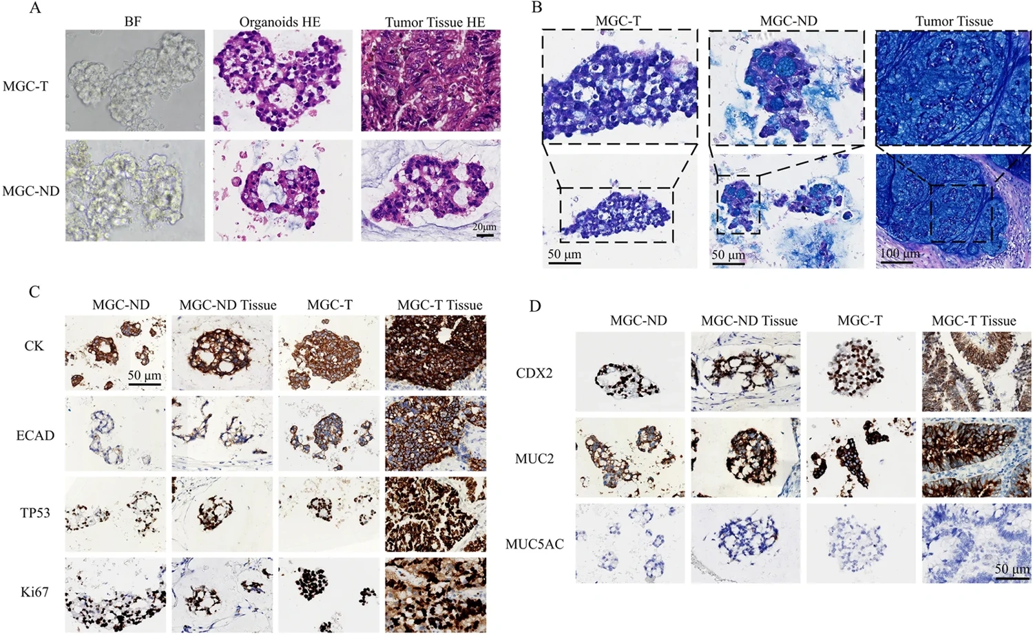
A team at the Seventh Affiliated Hospital of Sun Yat-sen University established organoids from mucinous gastric cancer, a histological subtype in which extracellular mucin is a prominent feature. Four models came from three patients. Two models, MGC-T and MGC-ND, represented a primary tumor and a peritoneal metastasis from the same patient; MGC2 and MGC3 came from two additional patients.
The organoids retained tumor-like morphology and expression of epithelial and gastric cancer markers. Alcian blue staining, mucin markers and live imaging allowed the team to follow mucin production as well as overall growth. Live imaging of MGC-ND captured vacuole formation and mucin release, followed by restoration of the cells' morphology.
A Chemotherapy Panel Reveals Different Response Patterns
The drug experiments used an 11-agent panel and measured ATP-associated viability with CellTiter-Glo 2.0 after 72 hours. The mucinous models were compared with six nonmucinous gastric cancer organoid models. The study used technical triplicates and three independent biological experiments for its concentration-response measurements.
The mucinous organoids showed limited sensitivity to fluorouracil under the tested conditions, with IC50 not reached at the highest concentration. Oxaliplatin responses varied even within the matched primary–metastasis pair: MGC-T did not reach an IC50 within the tested range, whereas MGC-ND had an IC50 of 5.23 μM. This difference illustrates why a shared histological subtype or patient origin does not ensure identical drug response.
Taxanes were active at lower concentrations in the four mucinous models. Paclitaxel IC50 values in MGC2 and MGC3 were approximately 0.0011 and 0.0016 μM, and docetaxel values were approximately 0.0007 and 0.0005 μM. The paired primary and metastatic models also showed sensitivity to these agents. These were experimental organoid results; the study did not test whether taxane selection improved patient outcomes.
A Small Clinical Comparison Used Mixed Histological Subtypes
Clinical follow-up was available for eight patients receiving oxaliplatin–fluorouracil-based treatment: six with nonmucinous disease and two with mucinous cancer. The first patient who supplied the paired MGC-T and MGC-ND cultures was lost to follow-up and excluded from this comparison.
The investigators classified organoid response using a normalized area-under-the-curve threshold and compared it with the clinical course over 12–36 months. Seven of the eight cases were concordant. The reported 87.5% agreement therefore describes this mixed gastric cancer group, rather than eight patients with mucinous gastric cancer. Early recurrence contributed to the definition of poor clinical response.
What the Model Adds
The study combines subtype-specific morphology, dynamic mucin behavior and pharmacological testing in patient-derived material. The matched primary and metastatic cultures add a within-patient comparison to the between-patient panel. Together, those features make the models useful for investigating why gastric tumors with similar pathology can respond differently to treatment.
The mucinous collection remained small, and its clinical observations did not validate the taxane findings. The drug response, mucin imaging and tumor-marker measurements address different aspects of the model. Extending all three across more patients would help establish how consistently the experimental behavior represents mucinous gastric cancer.
The authors identify a specific measurement limitation: CellTiter-Glo 2.0 is optimized for two-dimensional cultures, and mucin-rich, hydrogel-embedded organoids can impede reagent penetration and complete ATP release. Differences in lysis could affect viability estimates and apparent drug-resistance patterns. They therefore interpret the drug findings as qualitative trends requiring quantitative validation with a three-dimensional-culture-optimized assay.