Cartilage Organoid Screening Followed by Human Osteochondral Explant Testing
Miniaturized human cartilage pellets support dose/time screening of thyroid-receptor antagonists, followed by a native osteochondral tissue comparison.
Bone & CartilageOrganoids
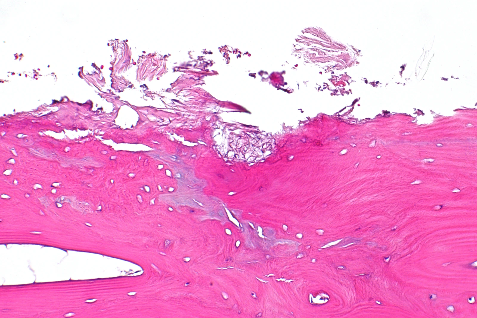
Femoral-head tissue from severe osteoarthritis, with cartilage loss. (Nephron, CC BY-SA 3.0)
Research Applications
Assess candidate effects on T3-induced terminal maturation in osteoarthritis-related human cartilage models.
Model & Design
Primary articular chondrocytes from preserved cartilage of joint-replacement patients form 96-well neo-cartilage organoids; osteochondral explants retain cartilage with underlying bone.
Experimental Format
T3-induced maturation followed by candidate compound dose/time comparisons; gene expression and secreted MMP3 are measured separately in each model.
Model Components
Component
Human material
Description
Macroscopically preserved cartilage from osteoarthritis surgical specimens; native explants and newly formed cartilage organoids are distinct tissue states.
Component
Perturbations
Description
T3 at 100 ng/mL; Galapagos-developed thyroid hormone receptor antagonists A, B and C. Organoid screening uses 0.3, 1, 3 and 10 μM for 4 or 7 days.
Component
Endpoints
Description
RT-qPCR for terminal/early maturation and cartilage-state markers; MMP3 ELISA in culture medium.
Measurements & Endpoints
CCDC80, ANKH and PHOSPHO1 expression
COL2A1, COL10A1 and ATOH8 expression
Secreted MMP3
Analysis
Evaluate markers within each tissue model, retain dose and duration distinctions and use the explant experiment to examine whether a screening response extends to native tissue.
Published Evidence
Study Design
Establish T3 responses in both systems, screen three compounds in organoids, and compare A/B responses in explants.
Study Sample
Dose/time screening: one donor with 1–2 samples per condition. Explant compound figure: 2–5 donors and 1–2 samples; these are different experiments from broader model-characterization sets.
Comparison
Untreated and T3-induced conditions, candidate doses/times and the two tissue formats.
Finding
Organoid Results report dose-dependent CCDC80 reduction, strongest at 10 μM/4 days. Compound A restored several terminal-maturation markers and MMP3 toward baseline in explants; B did not restore MMP3, while ATOH8/COL10A1 were unchanged.
Interpretation & Limits
The initial CCDC80 T3 rise was not significant in organoids (P = 0.27), unlike explants.
Explant B dose differs across Methods (10 μM) and Results/Fig.5 (20 μM).
Matrix-protein restoration and clinical efficacy were not established.
A 96-well cartilage organoid screen is followed by human osteochondral explant testing. Compound A most consistently reverses selected T3-related changes, while small screening samples and different marker responses constrain interpretation.