
Human Cartilage Models Compare Thyroid-Receptor Antagonists Across Tissue States
A 96-well cartilage organoid screen is followed by human osteochondral explant testing. Compound A most consistently reverses selected T3-related changes, while small screening samples and different marker responses constrain interpretation.
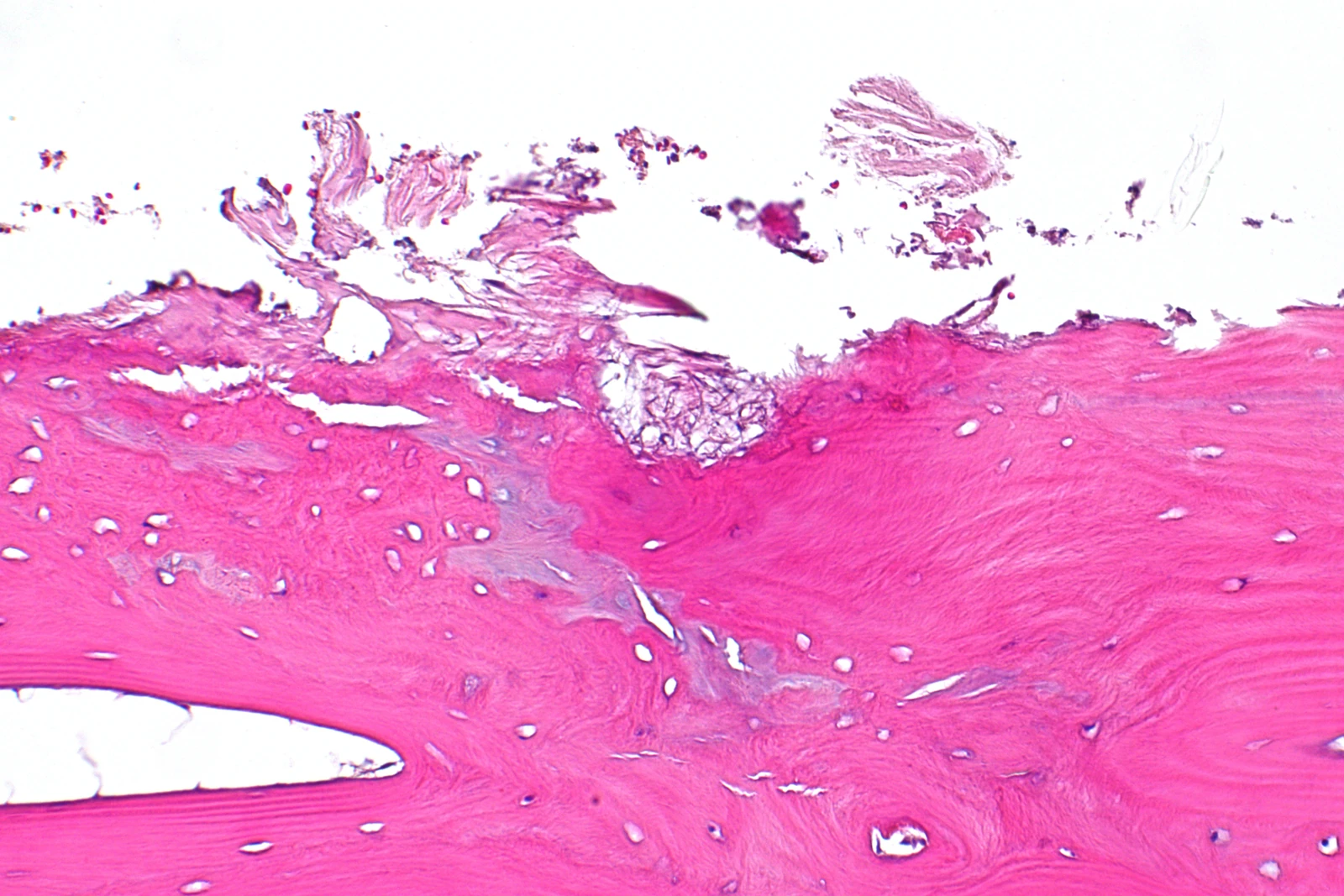
A Two-Stage Test for a Defined Cartilage Phenotype
A study from Leiden University Medical Center and collaborators combined a miniaturized cartilage organoid screen with a native human osteochondral explant comparison. The investigators tested compounds intended to block thyroid hormone receptor activation after inducing terminal chondrocyte maturation with triiodothyronine, or T3. The question was whether a response identified in newly formed cartilage would also appear in tissue retaining its original cartilage–bone organization.
Both systems used material from people undergoing joint replacement for osteoarthritis. Primary chondrocytes were obtained from macroscopically preserved cartilage and formed into neo-cartilage organoids in 96-well plates. The explants retained cartilage and underlying bone. These models therefore represented different tissue states from the same disease setting, rather than interchangeable versions of one assay.
T3 Responses Show Both Agreement and Differences
The organoid model was challenged with 100 ng/mL T3 for four days; explants received the same concentration for seven days. COL2A1 expression, used here as a marker of reactivation and matrix turnover, increased in both systems. ANKH, associated with terminal maturation and calcification, also increased in both.
CCDC80 responded more strongly in explants. Its organoid increase was modest and not statistically significant (P = 0.27), whereas the explant result was significant. COL10A1, an early-hypertrophy marker, decreased in organoids but did not change significantly in explants. These differences motivated using native tissue to follow up an initial screen, especially for markers affected by cartilage maturity.
The study also measured secreted matrix metalloproteinase 3 (MMP3). The authors interpret its recovery toward baseline as a matrix-turnover response within this T3-induced terminal-maturation setting. That interpretation is tied to these experimental conditions and does not make increased MMP3 a universal marker of cartilage protection.
Dose and Time Identify a Candidate for Explant Follow-Up
Galapagos developed and supplied three thyroid hormone receptor antagonists, designated compounds A, B and C. The organoid screen compared 0.3, 1, 3 and 10 μM over four or seven days. This dose/time experiment used cells from one donor, with one or two samples per condition, so it was a proof-of-concept screen rather than a multi-donor reproducibility study.
The Results report a dose-dependent reduction in CCDC80 expression for all three compounds, with the strongest effect at 10 μM after four days. Seven-day incubation did not provide an additional CCDC80 dose-response benefit. Compound A produced the most consistent MMP3 response across the tested conditions; B and C showed less consistent secretion patterns over the longer incubation.
Compounds A and B then entered the explant experiment. Compound A was tested at 10 and 20 μM, and the concentrations were pooled in the analysis after the authors reported no difference between them. The published Results and figure describe 20 μM B, whereas the Methods lists 10 μM, leaving that comparator dose uncertain. The compound-validation figure reports two to five donors, with one or two samples, distinct from the larger model-characterization experiments.
Marker Recovery Remains a Preclinical Tissue Result
In explants, compound A moved CCDC80, PHOSPHO1 and ANKH toward baseline and restored secreted MMP3. Compound B showed weaker gene-level effects and no comparable MMP3 recovery. Neither ATOH8 nor COL10A1 changed with the reported explant intervention. The unchanged earlier-stage markers help localize the response to selected terminal-maturation features.
The authors acknowledge that their principal evidence consists of gene expression and one secreted protein. They did not establish recovery of the cartilage matrix at the protein level, and technical difficulty prevented the intended matrix staining in the small organoids. The initial single-donor screen also needs broader donor testing. Systemic consequences of thyroid-receptor modulation and the feasibility of local treatment remain therapeutic-development questions.
The study positions neo-cartilage organoids for initial dose/time comparison, followed by native explant testing. The authors propose broader donor replication and possible use of induced pluripotent stem cell-derived chondrocytes for upscaling. Other osteoarthritis processes would require additional disease-relevant triggers and consideration of tissues such as synovium.