Exploratory assessment of brain-tumor drug delivery, exposure schedule and barrier injury.
Model & Design
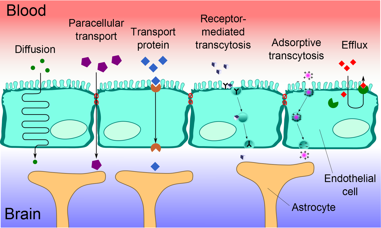
Human hCMEC/D3 endothelium with primary astrocytes and pericytes; two diffuse midline glioma models, SF8628 and KISPIDMG-105, formed into separate spheroids.
Experimental Format
32 plastic units per plate with tilt-driven perfusion, a porous membrane and a hanging-drop tumor compartment. Direct spheroid exposure, simultaneous barrier–tumor exposure and sequential permeate transfer are distinct designs.
Model Components
Component
Models and controls
Description
Preformed 500-cell tumor spheroids; barrier-cell spheroids for the direct screen; bare PET membrane and untreated cellular-barrier controls. At least two lots of primary supporting cells were used.
Component
Drug conditions and readouts
Description
Direct 24-hour cisplatin, doxorubicin, homoharringtonine and docetaxel screen. Chip follow-up used cisplatin/doxorubicin 0.5 or 1 μM and homoharringtonine 10 or 25 nM for 24 hours; ATP viability and fluorescent tracer permeability were measured.
Measurements & Endpoints
ATP-based viability and direct-exposure IC50
Lucifer Yellow and dextran barrier permeability
Response under coupled versus transferred-permeate exposure
Analysis
Compare tumor responses with a bare-membrane reference and with separate spheroids exposed to medium collected after 24-hour barrier permeation. Evaluate post-treatment tracer leakage separately from viability.
Published Evidence
Study Sample
Four to eight spheroids per well-plate condition; nine or eleven chip units per condition. Two tumor models.
Comparison
Untreated controls, acellular membrane, direct drug exposure and 24-hour permeation followed by a separate 24-hour tumor exposure.
Finding
The cellular barrier reduced apparent drug efficacy. KISPIDMG-105 viability (mean ± standard deviation) after 1 μM doxorubicin was 96.16% ± 8.16% with the barrier versus 64.83% ± 12.67% with bare membrane. Doxorubicin and homoharringtonine increased tracer permeability despite concentrations below barrier-spheroid IC50.
Interpretation & Limits
Transfer and continuous exposure have different concentration–time histories.
Drug concentrations were not directly quantified in each compartment; delivery limitation is functionally inferred.
Short exposure, simplified barrier and two tumor models lack clinical outcome or human PK validation.
Study Design
Primary study comparisons of the stated models, treatments and measurements.
A human-cell barrier coupled to two diffuse midline glioma models reduced the apparent activity of selected drugs. Simultaneous exposure and transfer of previously collected permeate produced different responses, while barrier leakage revealed injury missed by viability alone.