
A Perfused Blood–Brain Barrier–Glioma Chip Links Drug Delivery with Apparent Efficacy
A human-cell barrier coupled to two diffuse midline glioma models reduced the apparent activity of selected drugs. Simultaneous exposure and transfer of previously collected permeate produced different responses, while barrier leakage revealed injury missed by viability alone.
Connecting a Brain Barrier to Tumor Spheroids
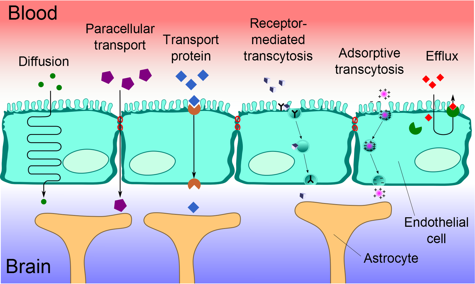
A drug that injures a brain-tumor culture must also reach the tumor through a vascular barrier. Researchers at ETH Zurich and University Children’s Hospital Zurich addressed both questions in a perfused platform reported in Microsystems & Nanoengineering. It couples a human-cell blood–brain barrier with diffuse midline glioma spheroids, allowing tumor response and barrier function to be measured under the same exposure conditions.
Each plastic plate contains 32 independent units. Human hCMEC/D3 endothelial cells form the vascular layer, supported by primary human astrocytes and pericytes across a porous membrane. An adjacent hanging-drop compartment holds the tumor spheroid. Asymmetric tilting circulates medium without separate pumps and tubing.
The two tumor models were the established SF8628 line and primary KISPIDMG-105 cells. Spheroids formed from 500 cells before transfer to the device. The team also checked whether a shared medium supported the different cell populations: it increased SF8628 viability relative to its usual medium, whereas the change in KISPIDMG-105 was not significant. This culture effect formed part of the assay background.
Direct Drug Sensitivity Provided the Starting Comparison
A 24-hour well-plate screen compared cisplatin, doxorubicin, homoharringtonine and docetaxel in tumor and barrier-cell spheroids. KISPIDMG-105 was sensitive to cisplatin, with an IC50 around 0.05 μM, while SF8628 showed little response over the tested range. Doxorubicin IC50 values were approximately 0.31 and 0.86 μM in the two tumor models, compared with 7.04 μM in barrier spheroids.
Homoharringtonine acted within a narrower concentration interval: its IC50 was about 20 nM in KISPIDMG-105, 3 nM in SF8628 and 40 nM in barrier spheroids. Docetaxel showed weaker activity under these conditions and was omitted from subsequent chip experiments. Well-plate conditions used four to eight spheroids per treatment; chip conditions used nine or eleven independent units.
Exposure Through the Barrier Weakened Tumor Responses
The chip experiments compared continuous barrier–tumor coculture with a bare-membrane reference. After 1 μM cisplatin, KISPIDMG-105 viability was 79.83% ± 10.24% with the cellular barrier and 58.69% ± 13.07% with the membrane alone. For 1 μM doxorubicin, the corresponding values were 96.16% ± 8.16% and 64.83% ± 12.67%. Values in these chip comparisons are means ± standard deviations (SD).
A separate design allowed drug to permeate the barrier for 24 hours, then exposed an independent tumor spheroid to the collected medium for another 24 hours. This accumulated permeate generally produced stronger effects than simultaneous coculture. SF8628 viability after 25 nM homoharringtonine was 96.51% ± 15.05% on the coupled chip, compared with 67.79% ± 11.04% after permeate transfer. These designs expose tumors to different concentration–time histories, even when the donor concentration is the same.
Barrier Function Added a Separate Safety Measurement
Before treatment, fluorescent tracer permeability was lower across the cellular barrier than across an acellular membrane. Tumor coculture slightly increased permeability to the smaller tracer. After treatment, 1 μM doxorubicin increased barrier permeability approximately twofold. At 25 nM, homoharringtonine increased permeability about fourfold for the larger dextran tracer and 1.8-fold for Lucifer Yellow. Cisplatin did not significantly disrupt the tracer barrier at the tested concentrations.
Leakage occurred at concentrations below the barrier-spheroid IC50. A cell-survival measurement therefore did not capture all functional injury. The study shows how barrier integrity, exposure timing and tumor sensitivity can give different information within one experiment. Its short exposures, two tumor models and simplified barrier do not reproduce patient pharmacokinetics; drug delivery was inferred from functional comparisons rather than measured drug concentrations in each compartment.