T-Cell-Integrated Human Skin Equivalents for Apremilast Response
Primary human skin equivalents with different CD4+ and CD8+ T-cell combinations support tissue and cytokine measurements during anti-inflammatory drug testing.
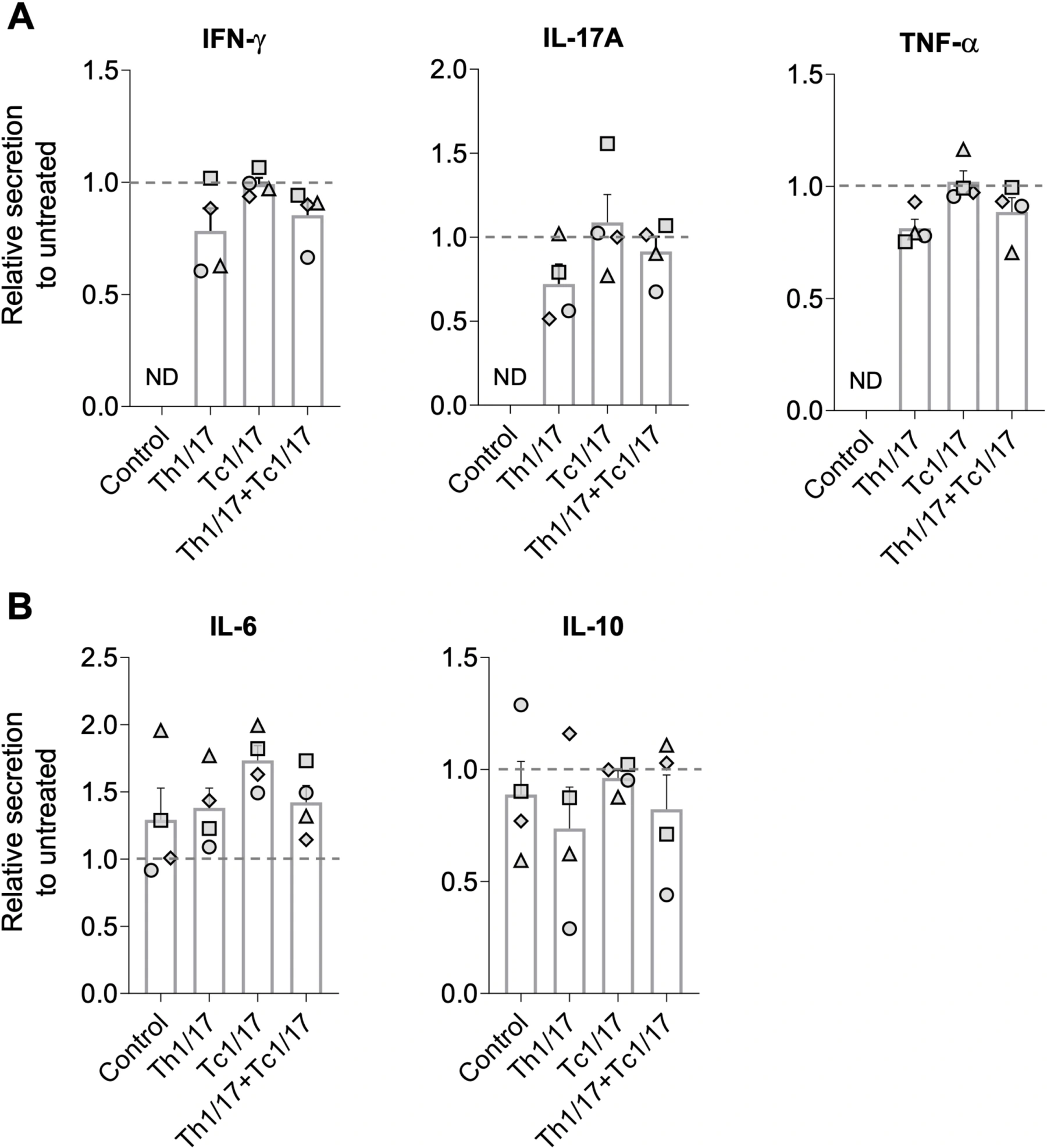
Retain donor-level patterns and distinguish independent donor experiments from within-experiment technical triplicates.
Published Evidence
Study Design
Compare subset composition with and without apremilast, alongside transferred T-cell-free controls and a non-transferred handling reference.
Study Sample
Four independent experiments using different skin/T-cell donors, with technical triplicates in each condition.
Comparison
Matched untreated cell combinations and T-cell-free skin equivalents.
Finding
CD4+ cultures showed cytokine-suppression trends in most donors without pooled statistical significance; CD8+ cultures were largely unresponsive. IL-6 increased across T-cell-containing donor cultures.
Interpretation & Limits
The small donor panel limits statistical power.
The immune/vascular composition is incomplete and current throughput is limited.
Healthy-donor reconstructed tissues reproduce selected psoriasis-associated features rather than a complete patient skin lesion.
Full-thickness human skin equivalents developed psoriasis-associated features after addition of defined CD4+ or CD8+ T-cell subsets. Apremilast responses differed by subset and donor, with cytokine-suppression trends in CD4+ cultures failing to reach statistical significance.