Cone-Selective Human Retinal Organoid Screening with Target and Duration Follow-Up
A 2,707-compound screen uses longitudinal cone imaging in one human iPSC background, followed by damaging/protective hit retesting, kinase profiling and selected genetic tests.
RetinaOrganoids
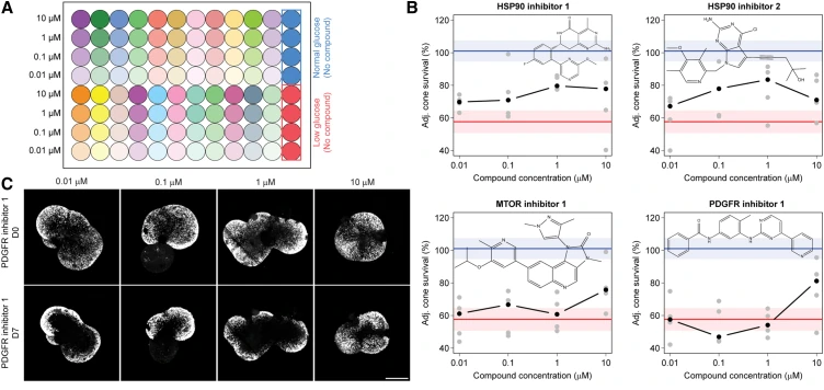
Figure 4A–C: secondary-screen layout, dose-response curves for four protective candidates and fluorescent retinal organoid images. The figure retains the compounds’ initial target labels. (Spirig SE et al., CC BY 4.0)
Research Applications
Identify photoreceptor-protective or damaging compounds while separating target annotation, causal evidence and delayed toxicity.
Model & Design
30-week retinal organoids from the human iPSC line 01F49i-N-B7; approximately 20,000 organoids overall and six independent productions in the primary screen.
Experimental Format
Low-glucose seven-day discovery with same-organoid baseline/follow-up cone counts; normal-glucose damaging-hit screen and low-glucose protective-hit screen; selected 14-day, rod, oxidative-stress and mouse follow-up.
Model Components
Component
Primary design
Description
2,707 known-target compounds at 10 μM, five replicate organoids each; approximately 15,000 organoids on 175 plates. Four normal- and four low-glucose 0.1% DMSO control wells per plate.
Component
Reporter validation
Description
Cone-specific reporter 97% ± 1% specificity and 64% ± 8% labeling efficiency, mean ± standard deviation, n = 5 each.
Component
Secondary design
Description
33 damaging candidates in normal glucose and 31 protective candidates in low glucose at 0.01, 0.1, 1 and 10 μM, with five replicates/condition.
Component
Mechanistic comparisons
Description
CS-KI-1/CS-KI-2 versus nonprotective structural analogs; 350-kinase profiling; additional CK1/MAPK11 inhibitors; cone-specific knockdown. 50 μM H2O2 supplies alternative injury.
Component
Cell-type expression comparison
Description
Bulk RNA sequencing of sorted cones and rods after seven days compares CS-KI-1/2 with their nonfunctional analogs and low-glucose controls, alongside HSP90 inhibition. Gene-set changes are interpreted as associations.
Measurements & Endpoints
Baseline-to-day 7 cone counts
Adjusted cone-survival score
Day 14 cone and rod survival
Kinase inhibition and cone-specific genetic response
Separate mouse retinal/outer nuclear layer thickness
Cone- and rod-specific transcriptomic responses
Analysis
The source covariance model adjusts for initial cone counts, with multiple-testing correction. Adjusted screening scores can exceed 100%; the smaller follow-up experiments use raw survival. Seven- and 14-day comparisons identify delayed changes in initially protective responses.
Published Evidence
Study Design
Five-replicate discovery/secondary screens with orthogonal chemistry, reporter checks, genetic and alternative-injury follow-up.
Study Sample
One human iPSC line; approximately 15,000 primary-screen organoids across six independent productions, with five replicate organoids per compound and secondary condition.
The primary screen identified 146 significantly damaging compounds; the first Results count reports 29 confirmations among 33 secondary retests. Four of 31 protective retests were significant. HSP90 inhibition protected cones at day 7 but was detrimental at day 14. CS-KI-1/2 protected cones through day 14; only CS-KI-2 retained significant day 14 rod protection. CK1-family knockdown improved cone survival; MAPK11 knockdown did not. Mouse CS-KI-1 outer nuclear layer preservation was nonsignificant (P = 0.069).
Interpretation & Limits
Metabolic and oxidative injury in one iPSC background provides a defined screening context; disease-genotype variation was not tested.
Initial mTOR/PDGFR annotations did not identify the protective mechanism; MAPK11 causal interpretation remains less secure than CK1.
The publication gives inconsistent totals of 28 and 29 confirmed damaging secondary compounds; the first dedicated result paragraph reports 29 of 33.
The narrative and parenthetical statistics give different doses for one MAPK11 inhibitor under hydrogen-peroxide injury, limiting dose-specific interpretation.
Mouse follow-up measures retinal structure; visual recovery was not assessed.
A 2,707-compound screen found protective and damaging responses in human retinal organoids. Follow-up implicated casein kinase 1, while HSP90 inhibition switched from short-term cone protection to harm at day 14 and a MAPK11 knockdown failed to improve cone survival.