
Human Retinal Organoid Screen Links CK1 Inhibition to Photoreceptor Survival
A 2,707-compound screen found protective and damaging responses in human retinal organoids. Follow-up implicated casein kinase 1, while HSP90 inhibition switched from short-term cone protection to harm at day 14 and a MAPK11 knockdown failed to improve cone survival.
A Cell-Specific Readout Made a Large Organoid Screen Possible
Researchers at the Institute of Molecular and Clinical Ophthalmology Basel and collaborating Novartis teams used human retinal organoids to find compounds that alter cone-photoreceptor survival. The Neuron study produced approximately 20,000 organoids and screened 2,707 compounds with annotated protein targets. Subsequent chemical, genetic and animal experiments tested whether the protective signals reflected the targets initially assigned to those compounds.
The organoids came from one human induced pluripotent stem cell line and matured for 30 weeks. A cone-selective fluorescent reporter labeled cells with 97% ± 1% specificity and 64% ± 8% labeling efficiency (mean ± standard deviation, n = 5 each). Automated imaging counted labeled cones in the same organoid before exposure and seven days later. Approximately 15,000 organoids, distributed across 175 plates and six independent organoid productions, supplied the primary screen.
Low-glucose medium induced approximately 40% cone loss over seven days, while normal-glucose controls retained their cone numbers over that period. Glucose became undetectable earlier in the low-glucose medium. This metabolic injury supplied a reproducible screening challenge, although organoids contacting the well bottom experienced additional spatial stress. It was not a patient-mutation model of retinal degeneration.
The Screen Detected Damage as Well as Protection
Each compound was tested at 10 μM in five replicate organoids, with vehicle controls in low and normal glucose. Because initial cone counts influenced later survival, the investigators adjusted the screening readout and used covariance analysis with correction for multiple comparisons. The adjusted score is a statistical quantity and can exceed 100%; it should not be read as a literal percentage of surviving cells.
The primary screen identified 146 compounds that significantly worsened cone loss. Of 33 selected damaging compounds retested in normal glucose at 0.01, 0.1, 1 and 10 μM, 29 again reduced cone numbers. Broad class I/II histone deacetylase inhibition was particularly harmful, whereas class III sirtuin inhibitors did not show the same pattern. Tubulin-targeting compounds also included damaging hits. Greater target breadth among class I/II inhibitors was associated with lower cone survival.
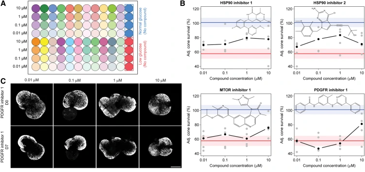
Seven compounds significantly protected cones in the primary screen. These and 24 other high-ranking candidates entered the protective secondary screen, where four of 31 showed significant benefit after multiple-testing correction. Two inhibited HSP90, while the other two were initially annotated as mTOR and PDGFR inhibitors. Target labels did not by themselves establish the mechanism of cone protection.
Longer Follow-Up Changed the Interpretation of a Positive Hit
An HSP90AA1 inhibitor at 1 μM improved cone survival at day 7 but became detrimental by day 14 compared with the low-glucose control. It did not significantly protect rods at the assessed times. The other two compounds, renamed CS-KI-1 and CS-KI-2, protected cones at 10 μM at both seven and 14 days. Both also improved rod survival at day 7, but only CS-KI-2 retained a significant rod benefit at day 14.
Several controls addressed the fluorescent readout. The protective compounds did not increase cone counts in normal glucose, and the fraction of labeled cells expressing the cone marker ARR3 remained comparable across conditions. Closely related chemical analogs that shared the original mTOR or PDGFR annotation did not protect cones. These comparisons argue against either reporter enhancement or the initial target annotation as sufficient explanations.
Profiling 350 human kinases identified differential inhibition of CSNK1G1, a casein kinase 1 (CK1) family member, by CS-KI-1, and MAPK11 by CS-KI-2. Additional CK1 inhibitors protected cones and rods at selected concentrations. Two of three tested MAPK11 inhibitors protected cones; the responses were not uniform across compounds or doses.
Bulk RNA sequencing of sorted cones and rods after seven days also distinguished the protective compounds from their nonfunctional analogs and low-glucose controls. HSP90 inhibition caused broad transcriptional shifts in both cell types. Changes specific to CS-KI-1 included downregulation of apoptosis-related gene sets in cones and rods and inflammatory-response programs in cones. CS-KI-2 was instead associated with increased cholesterol-homeostasis and mTORC1-signaling gene sets in both populations, with fatty-acid-metabolism programs also increased in cones. These expression associations suggest different responses to the compounds; they do not establish which pathway causes photoreceptor protection.
Genetic and Alternative-Injury Tests Narrowed the Target Evidence
Cone-specific knockdown of CSNK1G1 or CSNK1G3 improved cone survival, supporting a cell-intrinsic role for CK1-family signaling. Equivalent targeting of MAPK11 did not improve survival. The authors considered incomplete knockdown, effects mediated through other retinal cells and uncertainty about the target itself. The negative genetic result remains relevant despite the favorable responses to some MAPK11 inhibitors.
Selected CK1 and MAPK11 inhibitors also protected cones challenged with 50 μM hydrogen peroxide. In dark-reared rd10 mice, CS-KI-1, CS-KI-2 and a CK1 inhibitor preserved total retinal thickness between postnatal days 27 and 42. The paired-eye comparisons involved five, six and four mice, respectively. Outer nuclear layer preservation reached significance for CS-KI-2 and the CK1 inhibitor; the CS-KI-1 result did not reach significance (P = 0.069). These animal experiments assessed structure rather than restored visual performance.
The combined experiments make CK1 a stronger mechanistic candidate than a screen annotation alone would provide. The single human iPSC background, metabolic injury conditions and different short- and longer-term responses also define the next comparison: whether protection persists across disease genotypes and exposures without introducing delayed photoreceptor toxicity.