Investigation of persistent developmental responses to anesthetic exposure in human neural tissue.
Model & Design
Brain microphysiological spheroids derived from one human iPSC line, containing neural and glial lineages.
Experimental Format
Nominal gas-phase 2.4% sevoflurane for four hours at week 8, followed by four additional culture weeks; carrier-gas control.
Model Components
Component
Human microtissues
Description
iPSC-derived neural progenitors differentiated into multicellular bMPS cultures.
Component
Measurements
Description
Immunofluorescence, cell-marker ratios, myelin-associated process length, qPCR and mTOR-associated protein/phosphorylation measurements.
Measurements & Endpoints
Tissue cross-sectional area and apoptosis
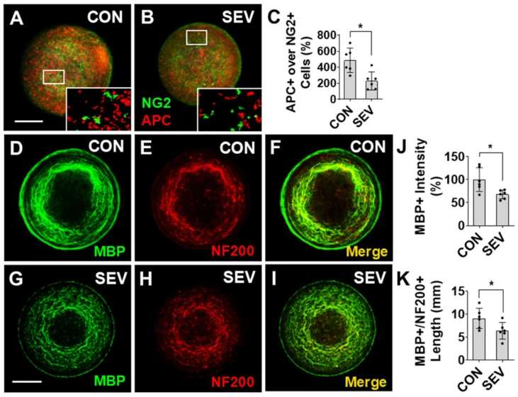
Neural and oligodendrocyte maturity-associated ratios
MBP/NF200-associated structures
Presynaptic marker density
mTOR-associated phosphorylation
Analysis
Interpret structural, expression and pathway-associated endpoints separately. Assay-specific cultures and differentiation batches remain distinct sampling units.
Published Evidence
Study Design
Compare delayed tissue phenotypes after sevoflurane versus carrier-gas exposure.
Study Sample
One iPSC line. Selected IF/WB cultures derive from two differentiation batches; vGlut1 qPCR uses six independent cultures.
Comparison
Carrier-gas controls under otherwise matched culture conditions.
Finding
Exposed cultures showed lower myelin-associated measurements and presynaptic markers and increased apoptosis and mTOR-associated phosphorylation. Some protein-intensity changes were not significant.
Interpretation & Limits
Gas stability and dissolved-medium exposure were not directly measured in this experiment.
Selected imaging/protein samples may include technical replication.
No electrophysiological synaptic validation or mTOR-rescue experiment was performed here.
A human brain microphysiological system showed lower myelin-associated measurements and presynaptic markers four weeks after sevoflurane exposure. The study combines neuronal, glial and pathway measurements within one iPSC background.