
Sevoflurane Exposure Is Followed by Persistent Developmental Changes in Human Brain Microtissues
A human brain microphysiological system showed lower myelin-associated measurements and presynaptic markers four weeks after sevoflurane exposure. The study combines neuronal, glial and pathway measurements within one iPSC background.
Measuring Development after an Exposure Interval
Johns Hopkins University researchers used a human brain microphysiological system to examine changes that persist after anesthetic exposure during development. The three-dimensional cultures contain neurons, astrocytes and oligodendrocyte-lineage cells, together with ongoing developmental processes. This provides a setting for studying drug-associated effects across interacting human neural and glial populations.
The tissues were derived from one human iPSC line and exposed at culture week 8 to a nominal gas concentration of 2.4% sevoflurane for four hours. Control cultures received carrier gas alone. The cultures then continued for another four weeks before the main tissue analyses. The selected exposure window coincided with ongoing neural development, synapse formation and myelination; it was not assigned an exact equivalent human age.
Changes in Tissue Size and Neural Populations
At week 12, the mean cross-sectional area was 0.17 mm² in exposed tissues and 0.20 mm² in controls. Cleaved-caspase-3-positive cell density was higher after sevoflurane. The ratio of MAP2-positive neurons to Nestin-positive neural progenitors was also lower, connecting the size change with altered cell-development measurements.
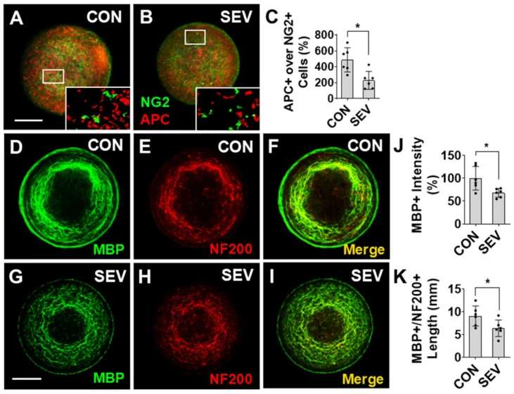
The study examined glial populations separately. The ratio of mature oligodendrocyte-marker-positive cells to precursor-marker-positive cells decreased. Myelin basic protein staining fell to approximately 68% of control intensity, and the total length of processes co-labeled for the axonal marker NF200 and myelin basic protein decreased.
These are marker-based and structural observations of oligodendrocyte development and myelin-associated structures. The authors note that the mature-to-precursor ratio can reflect several processes, including differentiation, survival and altered marker expression. The ratio alone does not isolate oligodendrocyte proliferation.
Synaptic Markers and mTOR-Associated Signaling
The density of synaptophysin-positive presynaptic puncta declined from approximately 6,679 to 4,298 per mm², and vGlut1 transcript abundance decreased. The postsynaptic GluN2B protein measurement showed a downward trend that was not statistically significant. GFAP-positive astrocyte density decreased, while the change in overall GFAP immunoreactivity did not reach statistical significance.
The researchers also measured signaling associated with mTOR. Phosphorylated S6 immunoreactivity and the phosphorylated-to-total p70S6K ratio increased. These measurements support a pathway-associated response, but this experiment did not test whether mTOR inhibition reverses the observed developmental phenotype.
Likewise, the presynaptic staining results were not accompanied by electrophysiological validation in this study. They identify changes in synapse-associated structures and markers without directly quantifying the resulting network function.
Exposure and Replication Define the Interpretation
The gas concentration describes the nominal exposure condition. Sevoflurane stability and dissolved-medium concentration were not directly measured in this experiment; the authors relied on a previously used delivery system. Measuring tissue-relevant exposure is one of the next steps they identify.
Replication also differed by assay. Immunofluorescence and Western blot comparisons drew on selected cultures from two differentiation batches, whereas the vGlut1 qPCR comparison used six independent cultures. The authors acknowledge possible technical replication within some imaging and protein measurements. These counts cannot be treated as six independent donor backgrounds.
The experiment adds a delayed-observation design to human developmental drug evaluation: a defined exposure is followed by a recovery interval and separate neuronal, glial, myelin and molecular measurements. The findings describe this culture system and exposure regimen. Additional cell lines, direct exposure measurement and functional neural assays would extend the evidence beyond its current scope.