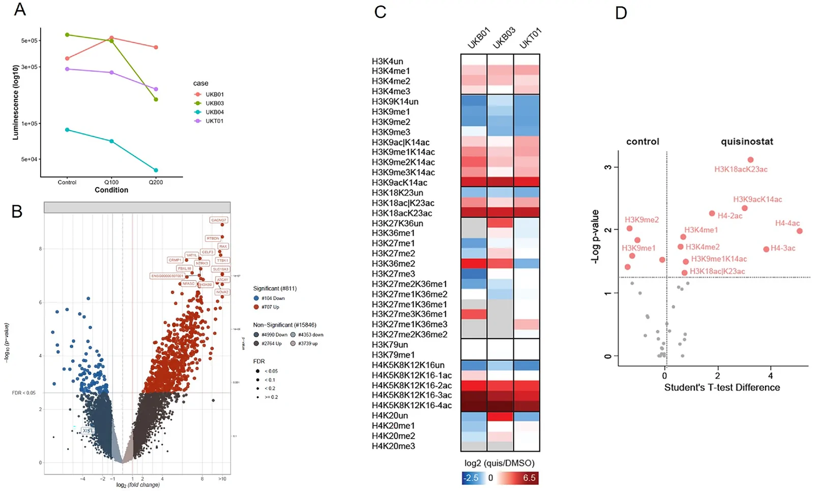
Ex vivo evaluation of epigenetic pharmacology and patient-specific model fidelity in uveal melanoma.
Model & Design
ALI organoids established from five patients; histone and drug experiments used their stated subsets.
Experimental Format
Matched tumor–organoid comparisons plus quisinostat, medium and DMSO conditions.
Model Components
Component
Source tissue
Description
Fresh or cryopreserved surgically resected uveal melanoma.
Component
Pharmacology
Description
Quisinostat at 100 nM and 200 nM for 72 hours, with matched medium/DMSO controls.
Measurements & Endpoints
ATP-associated viability luminescence
Differential gene expression
Mass-spectrometry histone H3/H4 modifications
Tumor morphology and molecular features
Analysis
Keep the five-patient establishment cohort, four matched histone pairs and two-tumor/eight-sample RNA drug experiment separate.
Published Evidence
Study Design
Matched tissue characterization combined with HDAC inhibitor treatment.
Study Sample
Five source patients; four matched tumor–organoid histone comparisons; two tumors/eight organoid samples for drug RNA profiling.
Comparison
Matched source tumor tissue and medium/DMSO treatment controls.
Finding
Quisinostat increased several H3/H4 acetylation marks and changed gene expression; viability varied, while bright-field morphology showed little difference.
Interpretation & Limits
Model establishment, molecular profiling and viability measurements used different subsets.
The cultures had limited expansion and lacked detectable immune/vascular components.
Air–liquid interface cultures preserved patient-specific features of uveal melanoma while showing culture-associated chromatin changes. Quisinostat altered histone acetylation and gene expression, with variable viability responses.