Investigation of early developmental drug effects on cell progression and the neural tissue microenvironment.
Model & Design
Human iPSC-derived dorsal forebrain organoids, with a separate cortical-organoid preparation for extracellular-matrix and stiffness follow-up.
Experimental Format
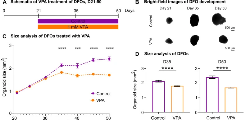
Initial valproic acid/levetiracetam dose comparison followed by 1 mM valproic acid from day 21 to day 50; imaging, RNA and protein analyses use defined but distinct sampling panels.
Model Components
Component
Human neural models
Description
Dorsal forebrain organoids from specified control iPSC lines and differentiation batches.
Component
Measurements
Description
Growth imaging, SOX2/Ki67 and neuronal staining, bulk/single-cell RNA sequencing, LC–MS secretome/proteome analysis, nanoindentation and second-harmonic generation.
Measurements & Endpoints
Organoid growth
Proliferative radial-glial proportion
Ventricular-like structure and cell-state composition
Secreted matrix/adhesion proteins
Tissue stiffness
Analysis
Compare each modality within its line/batch design and integrate biological-process patterns; gene/protein overlap is limited.
Published Evidence
Study Design
Compare valproic acid with control and an initial levetiracetam reference, then connect early developmental changes with extracellular measurements.
Study Sample
Main longitudinal imaging: four BIONi010-C differentiation batches. Protein experiments use separate line panels and pools of three organoids or media samples.
Valproic acid reduced growth and proliferative radial glia without increased vRG apoptosis at day 35. Later cultures had progenitor-rich states, matrix/adhesion changes and increased stiffness in the follow-up model.
Interpretation & Limits
The exposure window centers on early progenitors and neuronal differentiation.
Molecular, protein and imaging panels have different denominators.
A causal link between matrix accumulation and mechanosensing changes was not established.
Human forebrain organoids exposed to valproic acid showed slower growth, altered progenitor-to-neuron progression and changes in secreted matrix proteins. Transcriptomics, protein analysis and mechanical measurements examined different parts of that response.