
Valproic Acid Alters Forebrain Organoid Development and the Extracellular Environment
Human forebrain organoids exposed to valproic acid showed slower growth, altered progenitor-to-neuron progression and changes in secreted matrix proteins. Transcriptomics, protein analysis and mechanical measurements examined different parts of that response.
Following Early Neural Development during Drug Exposure
A German academic team used human induced pluripotent stem cell-derived dorsal forebrain organoids to investigate developmental effects of valproic acid. The study links changes in tissue growth and ventricular-like regions with cell-resolved transcription, secreted proteins and measurements of the extracellular environment. Its focus is the early progenitor and neuronal differentiation period.
An initial comparison tested valproic acid and levetiracetam at 0.1, 0.5 and 1 mM from differentiation day 21 to day 35. At 1 mM, valproic acid reduced organoid size and the proportion of proliferating radial glia. Levetiracetam produced no significant corresponding size or proliferative-radial-glia changes under the tested conditions. Apoptotic staining in the ventricular radial-glial compartment did not significantly increase with valproic acid at day 35.
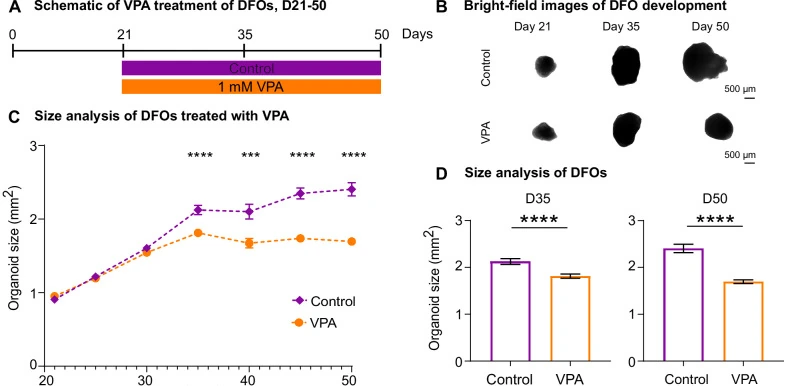
The investigators therefore followed 1 mM valproic acid exposure from day 21 to day 50. Treated organoids stopped growing around day 35 while control cultures continued to enlarge. Their ventricular-like structures became smaller or disorganized, and the proliferating radial-glial fraction was reduced.
Cell Composition Changes alongside Developmental Progression
Bulk RNA sequencing highlighted extracellular matrix, adhesion and synapse-associated changes. Single-cell analysis supplied a more specific view of the affected populations. Treated organoids contained a higher proportion of radial-glial and intermediate-progenitor transcriptional states, while controls contained more mature neuronal states.
This progenitor-rich composition occurred alongside reduced radial-glial proliferation. The authors interpret the combination as altered differentiation and developmental progression, rather than accelerated progenitor expansion. Immunostaining and transcript-defined populations did not agree on every intermediate-progenitor measurement, emphasizing that RNA-based cell states and individual protein markers describe different features.
The study used different line and batch panels for its imaging and molecular experiments. Four differentiation batches of the BIONi010-C line supported the main longitudinal growth and ventricular-region comparisons. These repeated cultures are distinct from donor diversity, and their denominators are not combined with those of the single-cell or protein experiments.
Secreted Proteins Reveal a Changing Microenvironment
The team analyzed organoids and their conditioned media at days 20, 35 and 50 using mass spectrometry. The extracellular protein comparison showed more extensive changes than the whole-organoid protein analysis. At day 50, 325 proteins increased and 547 decreased in abundance in treated-culture media relative to controls under the study’s statistical criteria.
Extracellular matrix, collagen organization and adhesion-associated proteins became prominent among the changes. Individual genes and proteins overlapped only modestly across the different assays. The investigators therefore compared shared biological-process patterns across data types, identifying matrix and adhesion modules that changed consistently.
A separate cortical organoid preparation tested the physical component of this response. Valproic-acid-treated tissues showed altered collagen organization, increased stiffness by nanoindentation and localized collagen-associated second-harmonic-generation signals. This follow-up supports a measurable change in the tissue environment in addition to a transcriptional signature.
An Exposure Model with Several Linked Endpoints
The study provides a framework for developmental drug evaluation that follows cell identity and tissue context together. Growth, progenitor behavior, neuronal differentiation, secreted proteins and stiffness describe related responses, each measured through a different experimental approach.
The exposure period captures early neurodevelopment and does not directly assess later synaptic activity or mature neural-network function. The authors also identify causal relationships between matrix accumulation and mechanosensing as a remaining experimental question. The observed extracellular and developmental changes therefore support further mechanistic investigation without establishing which one initiates the other.
For evaluating developmental effects, the useful feature is this combination of endpoints and time points. It shows how a drug-associated phenotype can involve developmental progression and the surrounding matrix even when increased apoptosis is absent from the initially examined progenitor compartment.