
Chamber-Like Cardiac Organoids Distinguish Functional and Structural Drug Effects
Human stem-cell-derived cardiac organoids formed stable chamber-like structures and registered changes in beating, chamber area and cell injury. Drug comparisons also showed that developmental exposure timing altered the measured response.
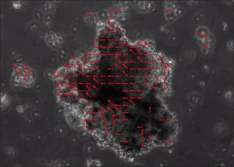
A Visible Chamber for Following Tissue Changes
A Westlake University-led team developed human pluripotent-stem-cell-derived cardiac organoids with a visible internal chamber. The model combines an outer myocardial region with an early endocardial-like layer, providing a setting in which shape and contraction can be followed together. The study examines developmental signaling and then uses several drugs to test the model's functional and structural responses.
The reported chamber-formation success rate was approximately 95% across eight independent experiments and more than 600 organoids. These numbers describe organoid generation rather than the sample size of every drug experiment. The structures could persist beyond 90 days, while molecular profiling indicated predominantly early developmental and atrial-like features.
The investigators examined FGF, WNT, BMP, retinoic-acid and activin-A signaling, along with transcription factors associated with chamber formation. The resulting architecture made brightfield chamber-area measurements possible without requiring destruction of the tissue at each observation point. Other assays supplied information about cellular composition, calcium signals and injury.
Separating Changes in Beating from Structural Damage
Isoproterenol provided a functional comparison. It increased beating frequency from 61.43 ± 2.34 to 87.26 ± 3.99 beats per minute in the reported experiment, with no detectable corresponding morphological change. Propranolol pretreatment suppressed spontaneous beating and prevented the increase after isoproterenol, supporting a β-adrenergic contribution to the response.
Doxorubicin produced a different phenotype. Chamber area declined progressively during exposure, accompanied by lower cell viability, greater LDH release and increased cardiac-stress and inflammatory transcripts. Daunorubicin also reduced chamber area in a separate morphology comparison. The different measurements distinguish a drug that primarily changes beating under the tested conditions from anthracycline-associated tissue injury.
These results demonstrate why chamber geometry, contraction and cell-damage measurements are complementary. A change in movement alone does not describe the same tissue response as progressive chamber collapse with loss of viability. The model allows those patterns to be compared within a common structural setting.
Developmental Timing Changes the Drug Phenotype
The developmental-toxicity experiments used thalidomide and acitretin, with aspirin as a reference control. Earlier exposures disrupted chamber area and beating, but the patterns differed. Thalidomide-treated organoids could initially form chambers and later lose them, whereas acitretin caused abnormal morphology at an earlier developmental stage.
The researchers also tested thalidomide after chamber formation, from day 15.5 to day 36.5. That later exposure did not significantly change chamber morphology or beating in the reported experiment. The comparison shows how the same drug can yield different observations when the model is exposed at different stages.
Gene-expression analyses identified overlapping and distinct responses to the two developmental toxicants. The authors caution that enrichment of a pathway bearing a neural annotation can reflect stress-responsive genes or small differences in tissue composition. Such pathway labels were not treated as evidence that the cardiac organoids had developed a functioning neural network.
Matching the Model with the Evaluation Question
The platform's visible architecture supports repeated measurement of chamber integrity, while video, calcium and molecular assays extend the assessment beyond morphology. It can therefore be used to examine a selected combination of developmental mechanisms, cardiac functional effects and structural injury.
The model retains early developmental characteristics and does not reproduce a complete mature heart. Cellular composition also changes over culture time, including the endothelial component. Those features are relevant when deciding which stage to test and which endpoint to interpret. The successful formation of a chamber is one aspect of model quality, alongside its cell identity and measured pharmacological behavior.
The strongest drug-related comparisons in this study are tied to those specific endpoints and exposure windows. Preserving their distinctions makes the organoids useful for studying why a compound changes function, disrupts tissue organization or interferes with an earlier developmental process.