
Transparent Electrodes Track Electrical Changes and Doxorubicin Injury in Human Heart Cells
A transparent microelectrode chip recorded drug-induced electrical changes while allowing imaging of the same human stem-cell-derived cardiomyocytes. Acute channel blockade altered electrical activity with little visible damage, whereas two-day doxorubicin exposure disrupted cell structure and viability.
Recording Through the Electrode
A study from Konkuk University combined extracellular electrical recordings and microscopy in human induced pluripotent stem cell-derived cardiomyocytes. The microelectrode array used indium tin oxide, or ITO, a transparent conducting material that allows cells on the recording surface to remain visible. The aim was to follow changes in electrical function alongside changes in cell condition during drug exposure.
The cardiomyocytes came from a single induced pluripotent stem cell line reprogrammed from BJ fibroblasts. At differentiation day 30, 97.06% expressed cardiac troponin T, and staining favored a ventricular-like identity. Cells were placed on fibronectin-coated electrodes and allowed to stabilize before measurement. The chip contained 16 wells with two electrodes per well.
Calcium-sensitive fluorescence showed activity around the electrodes, while field-potential recordings captured depolarization and repolarization. The investigators measured beat period, field-potential duration and signal amplitude. They also calculated a duration corrected for beat period using Fridericia’s formula. Keeping both the original and corrected duration became particularly important when a drug strongly slowed beating.
Two Acute Probes Produced Opposite Electrical Responses
The baseline electrophysiological analysis included 12 measurements. Mean beat period was approximately 1.498 seconds, field-potential duration 0.320 seconds, corrected duration 0.280 seconds and spike amplitude 1.712 mV. Calcium-transient measurements were reported separately with three replicates. These measurements characterized the cells before drug testing; they were not a clinical prediction dataset.
Acute experiments used the hERG potassium-channel blocker E-4031 and the L-type calcium-channel blocker nifedipine. For each concentration step, cells equilibrated for 17 minutes before a three-minute recording. Drug-response plots used five replicates. The 0.1% dimethyl sulfoxide vehicle changed the main parameters by less than 5%.
E-4031 prolonged corrected field-potential duration by approximately 18.8%, 54.6% and 125.8% at 0.003, 0.01 and 0.03 μM, respectively. Nifedipine shortened it by approximately 26.3% at 0.08 μM and 62.4% at 0.8 μM. Nifedipine did not significantly alter signal amplitude. Despite these electrical effects, imaging after the highest acute concentrations showed no significant change in overall cell morphology.
Longer Doxorubicin Exposure Linked Electrical and Structural Injury
The longer experiment applied 3 μM doxorubicin and recorded the cells before exposure and after one and two days. Both treated and vehicle-control groups were followed over time, with five replicates in the electrophysiological comparison. Live/dead staining and sarcomeric imaging added information about the cellular changes accompanying the electrical signals.
Mean beat period rose to 8.213 seconds after one day of doxorubicin, compared with 1.961 seconds in controls. At day two, the corresponding values were 4.177 and 1.678 seconds. Thus, beating remained slower than in controls even though the greatest slowing occurred at the first observation.
Uncorrected field-potential duration increased from approximately 0.382 seconds before treatment to 0.448 and 0.495 seconds on days one and two. Corrected duration decreased sharply on day one and partly recovered on day two because its calculation also incorporated the large change in beat period. Signal amplitude fell from day one onward.
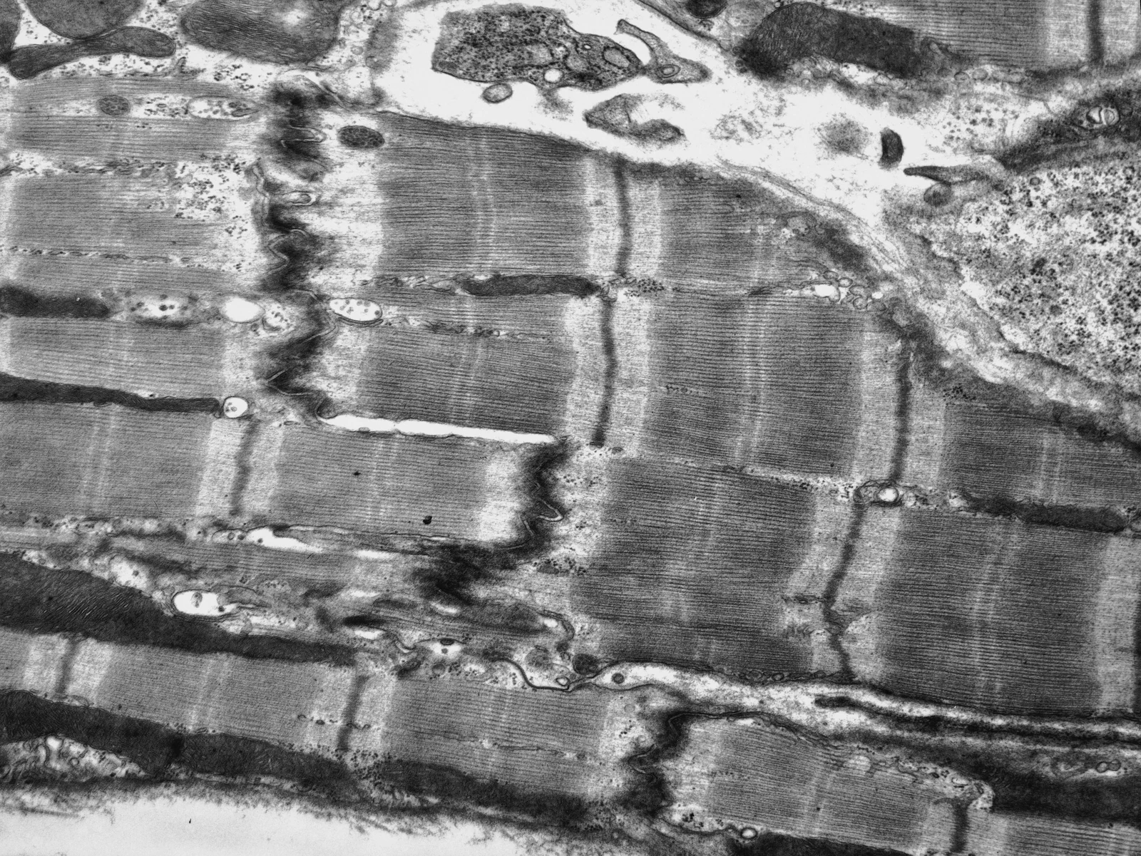
The optical findings supplied a complementary injury readout: cell density and viability declined, dead-cell staining increased and α-actinin staining showed disrupted sarcomeres. These changes contrasted with the limited morphological effects of the acute channel blockers. The comparison therefore showed why electrical and structural measurements can describe different stages or types of drug response.
The study tested three compounds in cardiomyocytes from one stem-cell source. Its two-dimensional cells retained the immaturity typical of this model, and the experiments did not establish performance across a large panel of clinically classified drugs. The authors identify maturation, three-dimensional tissue organization and additional contractility measurements as specific next steps for extending the assay.