
Vascularized Skin Assembloids Respond to Two Atopic Dermatitis Therapies
Human skin and blood-vessel organoids were assembled with immune cells to study allergen-associated inflammation. Upadacitinib and dupilumab improved barrier-related markers and reduced STAT6-associated inflammatory signals.
Combining Skin, Vascular and Immune Compartments
A Seoul National University-led study assembled human skin organoids and blood-vessel organoids into a vascularized skin model for inflammatory drug research. Adding cord-blood-derived mononuclear cells supplied an immune compartment. The resulting system was used to compare responses to the atopic dermatitis therapies upadacitinib and dupilumab.
The tissue modules were generated from human induced pluripotent stem cells and combined under air–liquid-interface conditions. The assembloids developed a stratified epidermis, a fibroblast-rich dermal region and interconnected vascular-like structures. These components could be examined in relation to barrier organization and the spatial distribution of immune cells.
The vascular compartment remained part of a static culture. Its endothelial junctions and supporting structures indicated vascular organization, while sustained physiological blood flow and a mature artery–vein–capillary hierarchy were outside the model. This distinction helps define which tissue–immune interactions the platform can investigate.
Allergen Exposure Produces an Inflammatory Tissue State
House dust mite extract triggered epidermal thickening, reduced barrier-associated filaggrin and increased keratinocyte stress markers. Immune cells accumulated in the dermal compartment, including near vascular structures. Transcriptional and protein measurements identified type 2-associated inflammatory mediators, including IL-4, IL-13, TSLP, CCL17 and CCL26.
Comparison with published atopic dermatitis skin-expression data found partial overlap in inflammatory and epithelial-stress programs. The experimental model therefore captures selected disease-associated responses following an allergen challenge, with tissue architecture and immune localization available alongside bulk molecular measurements.
The investigators used the barrier marker filaggrin and inflammatory mediators to choose drug concentrations. Upadacitinib responses improved up to 1 μM, while higher concentrations lost that advantage and produced rebound inflammatory-marker changes. Dupilumab responses approached saturation above 10 μg/mL. The dose-selection experiments supplied context for the subsequent tissue comparisons.
Barrier and Immune Measurements after Treatment
Both drugs reduced allergen-associated epidermal thickening and increased filaggrin, loricrin and claudin 1 expression. TSLP and the stress-associated keratin KRT16 decreased. These endpoints combined tissue morphology with proteins related to epidermal differentiation and junctions.
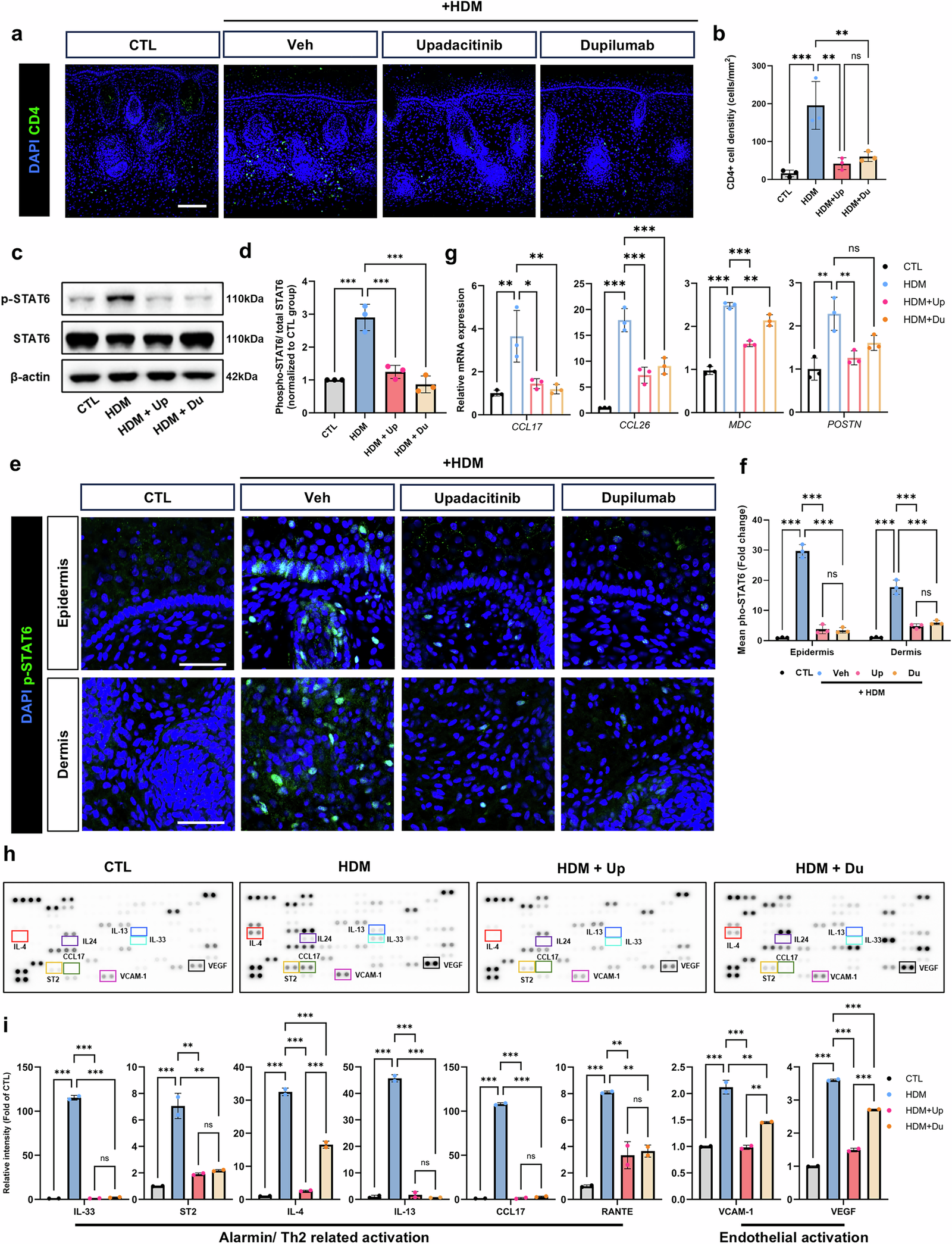
The immune comparison showed fewer CD4+ T cells in the upper dermis after treatment. Allergen-exposed tissues had increased phosphorylated STAT6 with little change in total STAT6. Upadacitinib and dupilumab reduced that phosphorylation response and its nuclear staining in epidermal and dermal compartments.
STAT6-associated target-gene and cytokine measurements moved in the same general direction. The drugs reduced selected chemokines, epithelial alarmins and vascular-activation-associated signals. These observations are consistent with JAK1 inhibition by upadacitinib and IL-4 receptor α blockade by dupilumab, which acts on IL-4/IL-13 signaling.
A Controlled Pharmacology Model of Selected Disease Features
The study links drug response with barrier proteins, immune-cell position and pathway-associated measurements in an organized human tissue. Its modular design allows skin and vascular components to be generated separately before immune incorporation and inflammatory challenge.
The immune cells come from cord blood and do not reproduce the complete resident immune repertoire of adult skin. The model also lacks the chronic recurrence and patient diversity of clinical atopic dermatitis. The reported comparisons establish responses to two selected therapies, rather than broad predictive performance across patients and drug mechanisms.
Within the experimental scope, the model provides a way to connect anti-inflammatory pharmacology with changes across epidermal, dermal and immune-associated compartments.