
Dequalinium Preferentially Kills Quiescent Cancer Cells Across Three Model Formats
Low-oxygen, nutrient-limited tumor models were more sensitive to dequalinium than actively growing counterparts. Detailed H2228 experiments linked the response to oxidative stress and ferroptotic cell death.
Comparing Cell State Within Each Model
A Japanese academic team tested whether quiescent cancer cells have a redox vulnerability that can be targeted pharmacologically. The study compared actively growing and growth-restricted versions of human H2228 lung cancer cells, H2228 multicellular spheroids and the C45 colorectal cancer organoid line. Human WI-38 lung fibroblasts provided an additional toxicity comparison.
Low oxygen combined with reduced serum or removal of growth supplements produced the quiescent conditions appropriate to each model. All three tumor formats grew under their general-growth conditions and showed little growth under the restricted conditions. This design allowed drug response to be compared between cell states within a model while retaining the distinctions between monolayers, spheroids and organoids.
A State-Dependent Difference in Drug Potency
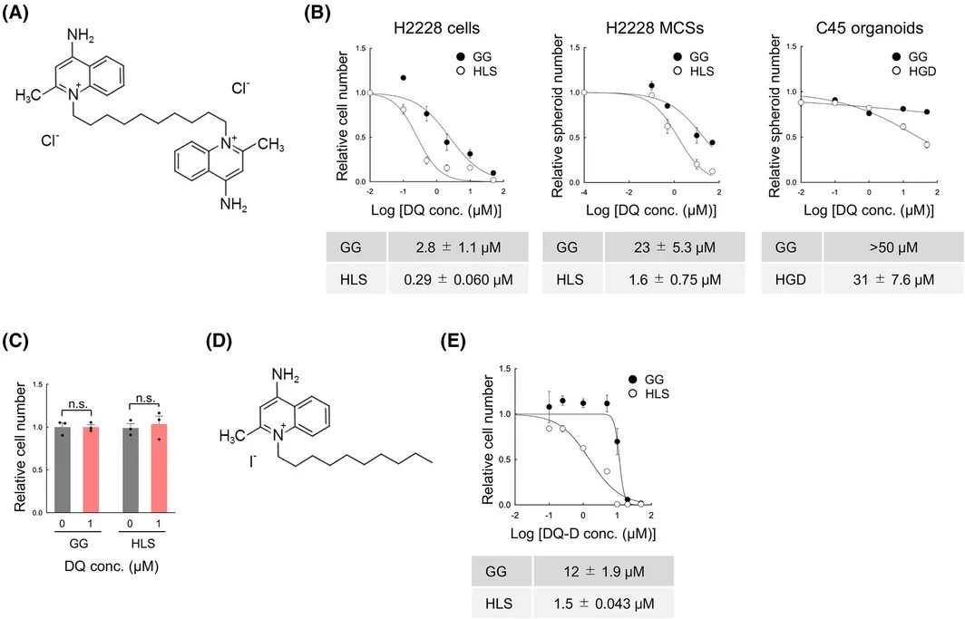
Dequalinium chloride was more potent in the quiescent conditions. The half-maximal effective concentration in H2228 monolayers was 0.29 ± 0.060 μM in quiescent cells and 2.8 ± 1.1 μM in growing cells. The corresponding values were 1.6 ± 0.75 μM and 23 ± 5.3 μM for H2228 spheroids, and 31 ± 7.6 μM and greater than 50 μM for C45 organoids. The study's concentration-response experiment reported five measurements per curve.
The investigators measured live-cell counts in the monolayer assay and counted spheroids or organoids in the three-dimensional assays. The numerical responses therefore remain tied to the particular model and measurement. WI-38 fibroblasts showed little toxicity in the tested comparison, adding a nonmalignant human-cell reference to the tumor-model results.
Mitochondrial Effects and Oxidative Stress
The more detailed mechanism experiments were performed in H2228 cells. Dequalinium reduced mitochondrial membrane potential and intracellular ATP in both growing and quiescent cells to a similar degree. Those effects did not explain the difference in killing. Quiescent cells already had higher reactive oxygen species levels and a higher ratio of oxidized to reduced glutathione before treatment.
After dequalinium exposure, reactive oxygen species and the glutathione ratio rose strongly in quiescent cells, with smaller changes in the proliferating cells. Rotenone provided a contrasting mitochondrial perturbation: it increased reactive oxygen species more strongly in growing H2228 cells and was more toxic in that state. The comparison linked the pattern of oxidative stress with the direction of cell-state selectivity.
Evidence for Different Forms of Cell Death
Quiescent H2228 cells accumulated lipid peroxides after treatment. Ferrostatin-1 prevented both lipid-peroxide accumulation and the associated cell death, supporting a ferroptotic mechanism in this setting. Growing cells instead showed a more limited, caspase-independent apoptotic response accompanied by movement of EndoG into the nucleus.
Expression of GPX4 and HMOX1 increased as part of the measured stress response. The intracellular labile iron pool did not increase significantly under the tested conditions or after drug treatment.
For model-based drug research, the study illustrates how a candidate's apparent activity can depend on the physiological state being tested. The C45 organoid result extends the state-dependent potency comparison to a patient-derived tumor model. The detailed ferroptosis evidence belongs to the H2228 experiments; the study did not establish an equivalent mechanistic dataset for every model or test dequalinium against quiescent tumors in vivo.