
Direction-Aware Drug Screening Leads to a HepG2 Test of a Two-Molecule Formulation
Independent direction and affinity models prioritize complementary molecules for liver-injury research. A coassembled hesperidin–ursodeoxycholic acid formulation improves selected HepG2 responses, while cold-start prediction and mechanistic coverage remain limited.
Predicting an Effect Requires More Than Binding Affinity
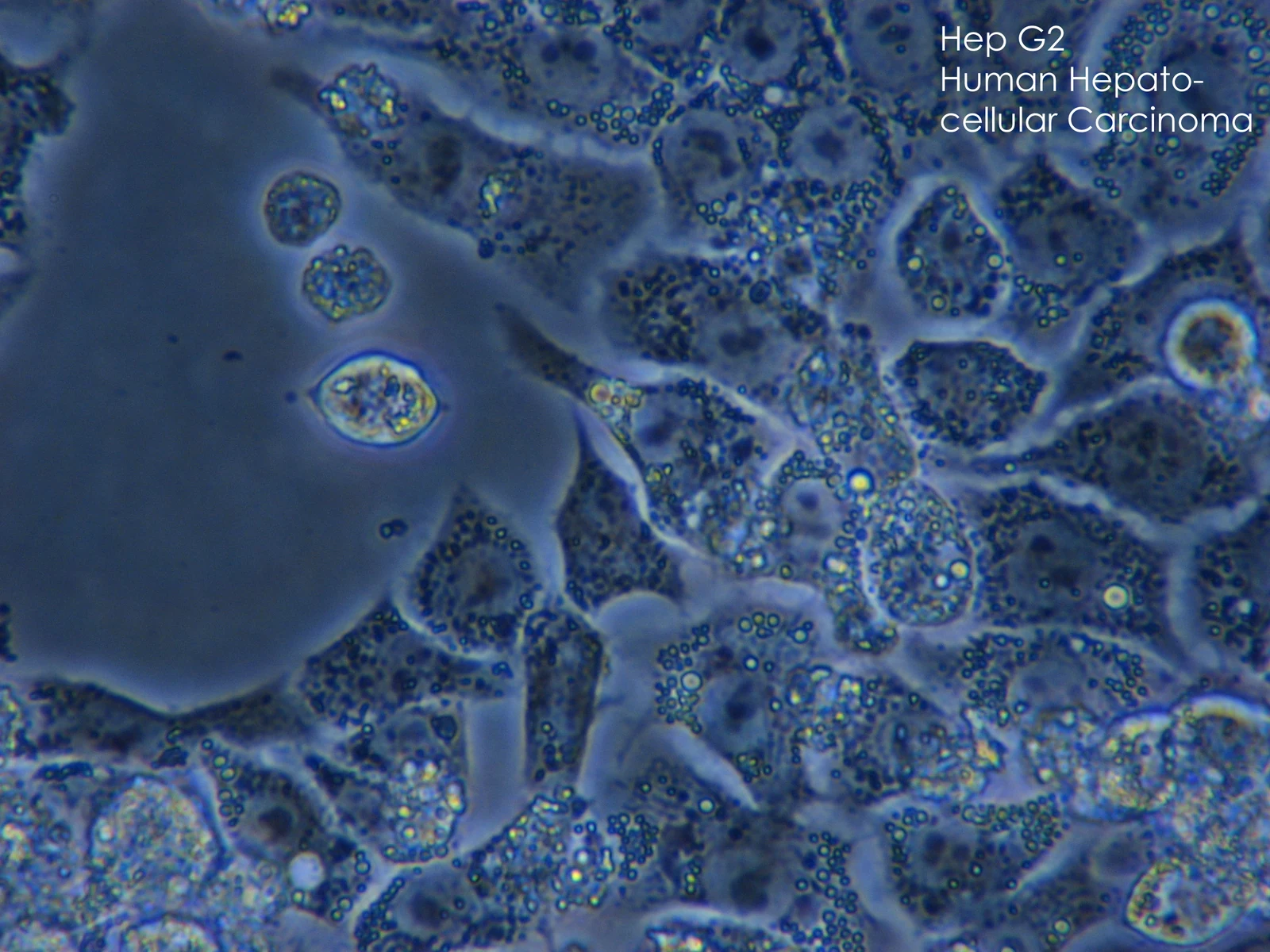
An academic and clinical research team in China developed a computational screening approach that considered both molecule–protein affinity and the intended direction of biological activity. The study linked a disease-network analysis with candidate selection, formulation development and cellular testing. Experiments in HepG2, a human liver-cancer cell line, provided one functional test of a selected hesperidin–ursodeoxycholic acid combination.
The computational component used two independently trained models with a shared type of architecture: one classified activating versus inhibitory annotations, and the other predicted affinity. The direction dataset contained 23,204 molecule–protein pairs, while the affinity dataset contained approximately 2.18 million pairs. These were separate tasks and training sets, rather than one combined pool of equally labeled observations.
Generalization Tests and Rat Transcriptomes Shape the Screen
Five architectures were compared under the same training settings. Performance was assessed with tenfold cross-validation under random pair splitting, an unseen-protein setting based on sequence grouping, and an unseen molecular-scaffold setting. Every architecture performed best under random warm-start conditions and lost performance in the two cold-start settings. The selected GSEC–GESM architecture combined a graph-based molecular encoder with a protein representation and offered the reported balance of performance and stability.
The biological screening directions came from a reanalysis of Open TG-GATEs rat liver transcriptomes after high-dose acetaminophen exposure. Profiles at 3, 6, 9 and 24 hours were compared with time-matched controls. The 24-hour profiles showed lower expression of cytokine-associated genes and increased glutathione-metabolism genes. The authors interpreted this as a repair-associated state and used suppression of the first pathway group and activation of the second as complementary screening objectives.
More than 10,000 bioactive compounds were evaluated computationally. Candidates from the two directions were then paired and tested for coassembly. Hesperidin and ursodeoxycholic acid were selected because their combination formed relatively small, uniform particles under the tested formulation conditions. This choice incorporated physical compatibility alongside predicted biological activity.
Formulation Measurements Add a Delivery Variable
The selected hesperidin–ursodeoxycholic acid nanomedicine (HUNMs) had a reported hydrodynamic diameter of 58.17 ± 1.76 nm and a polydispersity index of 0.104 ± 0.021. Hesperidin and ursodeoxycholic acid accounted for 42.4% and 57.6% of active-molecule content. Approximately 50% of both components was released within 24 hours in the reported buffer experiment.
Three independent molecular-dynamics simulations supported aggregation driven primarily by van der Waals and hydrophobic interactions, with surface–water hydrogen bonding contributing to dispersion. These calculations explained formulation behavior; they did not establish that every predicted disease target was directly engaged by the resulting particles.
In HepG2 cells, fluorescently labeled formulation produced greater intracellular signal after two hours than the free comparison. The study found no appreciable formulation-associated cytotoxicity in its 24-hour assay. Improved uptake also means that subsequent biological differences can reflect delivery and exposure, alongside the pharmacological properties of the two molecules.
Human-Cell and Animal Evidence Answer Different Questions
For the liver-injury comparison, HepG2 cells received 5 mM acetaminophen for two hours, followed by 24-hour treatment with free hesperidin, free ursodeoxycholic acid, their physical mixture or HUNMs. Treatments used an equivalent hesperidin concentration of 42.4 μg/mL. The coassembled formulation improved cell viability and glutathione peroxidase activity more than the free combination.
An additional oxidative-stress assay in HepG2 cells showed reduced intracellular ROS. Anti-inflammatory TNF-α measurements came from a separate mouse RAW264.7 macrophage model. Whole-animal testing used male mice, with six animals per group and outcomes assessed 24 hours after acetaminophen exposure; the formulation reduced injury-related biochemical and histological measurements. These results span different species, cell identities and experimental levels.
Prediction performance declined for unseen proteins and molecular scaffolds. The subsequent cellular comparisons used a rat-transcriptome-derived target hypothesis and one human tumor-derived cell line. Direct binding or mediation was not measured for every predicted target, while greater cellular uptake offers an additional explanation for the formulation’s stronger effects.