
Matched Immune Cells Expose Variable Checkpoint Responses in Head and Neck Cancer Organoids
Head and neck cancer organoids were paired with blood-derived immune cells to compare PD-1 and LAG-3 blockade. Responses varied by patient, and combined blockade did not consistently overcome resistance.
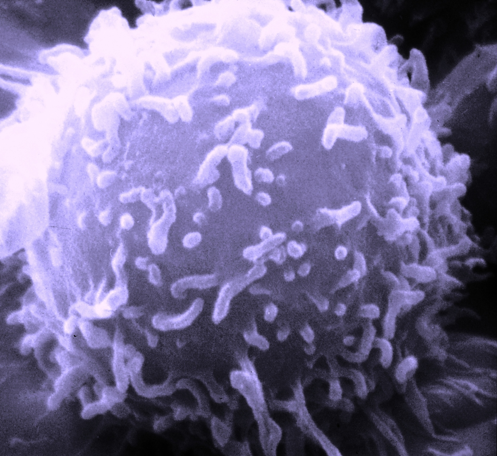
Pairing Tumor Tissue with the Patient’s Immune Cells
A German academic team led from Rostock established head and neck squamous cell carcinoma organoids and combined them with blood-derived immune cells. The aim was to examine patient-specific responses to immune checkpoint inhibitors in a system containing both tumor material and an immune effector population.
The team collected 38 tumor specimens from 35 patients, including fresh and biobanked samples. Twenty-eight specimens produced viable organoids, an overall success rate of 73.7%. Histological comparisons supported preservation of tumor architecture and epithelial markers, although immune cells originally present in the tissue declined during prolonged culture.
The assay therefore added peripheral blood mononuclear cells. Autologous cells from the same patient were compared with allogeneic cells from healthy donors where available. T-cell expansion was greater in the autologous setting, with mean expansion ratios of 2.3 and 1.3, respectively. Tumor viability and T-cell measurements used different evaluable subsets.
Testing PD-1 and LAG-3 Blockade
After a priming and expansion phase, the investigators compared nivolumab, pembrolizumab, relatlimab and nivolumab–relatlimab in a three-day effector assay. Calcein fluorescence measured change in organoid viability, while flow cytometry and secreted cytokines described immune responses. A reduction of at least 20% from baseline defined sensitivity for the study's binary classification.
Eleven cases had evaluable viability results. Using that threshold, 45.5% were sensitive to nivolumab, 36.4% to pembrolizumab and 27.3% to relatlimab. Some cultures responded broadly, while others remained resistant across agents. Combining nivolumab with relatlimab did not consistently increase tumor killing or overcome resistance.
Immune measurements showed that activation and exhaustion-associated markers could coexist. LAG-3 blockade increased several activation markers, while LAG-3, CTLA-4 or TOX expression persisted in relevant T-cell populations. A stronger activation signature did not translate into uniformly stronger organoid killing.
IL-17A Emerges as a Candidate Associated Signal
IL-17A and Fas ligand increased together in several cocultures. Higher IL-17A levels were associated with lower organoid viability after some checkpoint treatments. The team used two contrasting clinical cases to examine these patterns in more detail: a nivolumab responder showed coordinated cytokine and T-cell activation, whereas a pembrolizumab nonresponder showed limited activation.
Separate clinical analyses examined circulating cytokines. In 76 treatment-naïve patients, higher IL-17A was associated with better overall survival. Longitudinal monitoring in five patients receiving PD-1 blockade suggested an increase associated with stable disease. These clinical observations used different populations and measurements from the organoid assay.
What the Coculture Captures
The model preserves a matched tumor–blood relationship while allowing several checkpoint treatments to be tested against the same patient-derived tumor material. It can connect a viability change with T-cell phenotype and secreted mediators. The heterogeneous results are useful for studying why the presence of a checkpoint target or an activation signal does not ensure the same functional response in every culture.
Peripheral blood cells do not reproduce all tissue-resident immune populations or their spatial history. The study was small and largely retrospective, with limited molecular and dynamic profiling. Its IL-17A findings identify an associated candidate signal for further work; direct functional tests and larger prospective clinical comparisons are needed to establish its contribution to treatment response.