
Atovaquone Combination Activates T Cells in Head and Neck Cancer Organoids
Radiotherapy, atovaquone and PD-1/PD-L1 blockade increased CD8-positive T-cell activation and tumor killing in patient-derived head and neck cancer organoids. Separate mouse experiments showed substantial differences among tumor models.
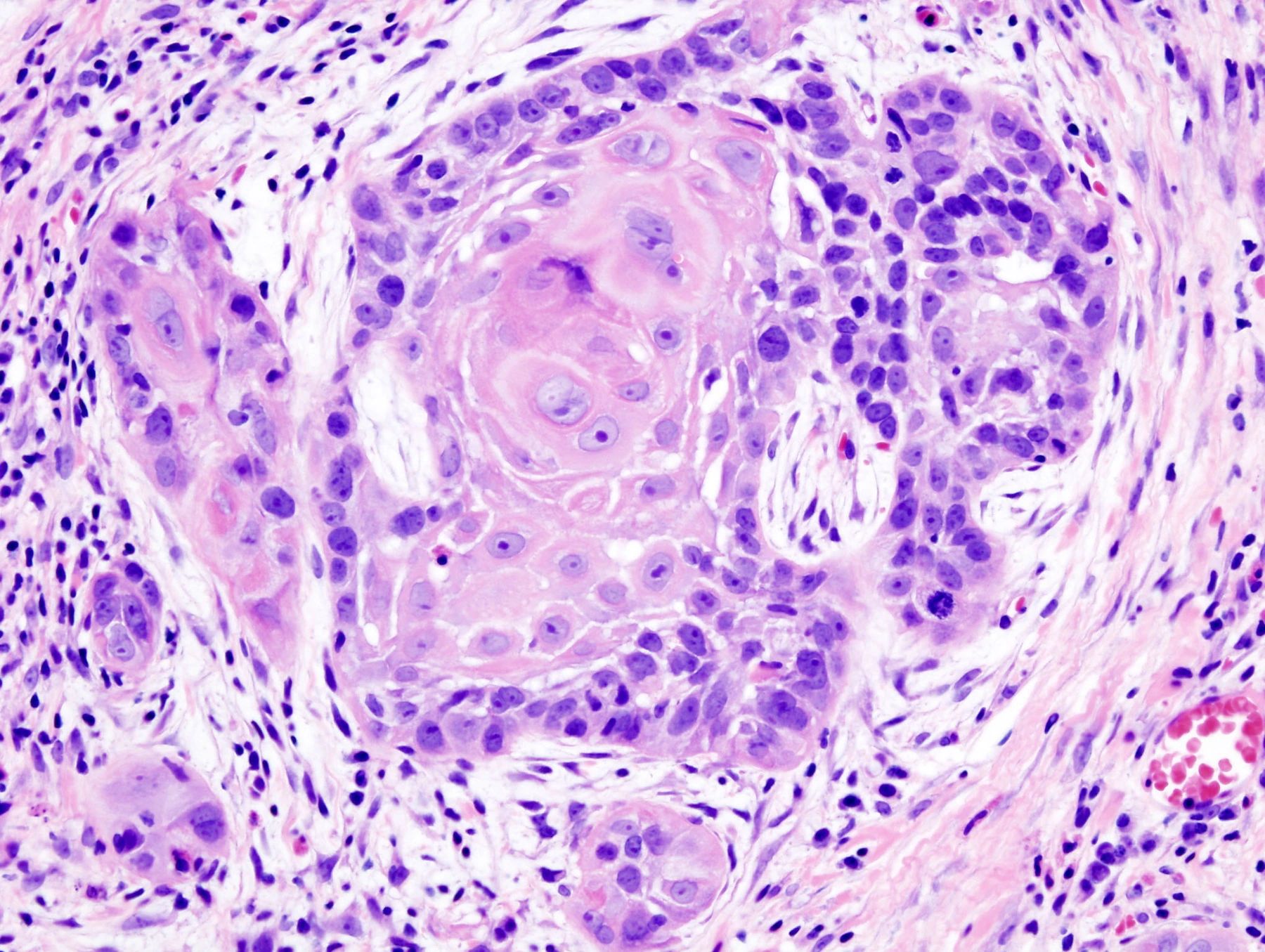
A Human Model for a Three-Part Treatment
Researchers at King's College London and collaborating institutions tested the antimalarial drug atovaquone alongside radiotherapy and immune checkpoint blockade in head and neck squamous cell carcinoma. The study combined mouse tumor experiments with patient-derived tumor organoids, allowing the investigators to examine immune activity in human-derived material as well as tumor growth in animals.
In the organoid experiments, treatment with radiotherapy, atovaquone and PD-1 or PD-L1 blockade increased CD8-positive T-cell activation and enhanced tumor-cell killing. This human-model result concerned the interaction between treatment and immune-mediated killing; the reported complete tumor eradications came from the separate animal experiments.
Responses Differed Among the Mouse Models
The triple combination delayed tumor growth in three syngeneic mouse models, with different degrees of response. In the MOCL1 model, tumors were completely eradicated in 6 of 12 mice. The other two models did not show complete eradication. Treated tumors also showed changes in cytotoxic T cells and several immunosuppressive cell populations.
Atovaquone reduced EGFR signaling and PD-L1 expression in the investigated systems. The authors also linked the radiotherapy–atovaquone combination to G2/M cell-cycle arrest and reduced homologous-recombination DNA repair. These findings provide several explanations to investigate for the variable responses. The organoid arm adds a patient-derived setting in which to study the immune component of the combination before any clinical evaluation.