
Kidney-Chip Materials Change Ischemia Modeling and CDDO Responses
Matched-geometry chips made from PET or PDMS produced different injury patterns under the same nominal oxygen conditions. Simulations and human proximal-tubule experiments linked platform oxygen transport with the observed drug response.
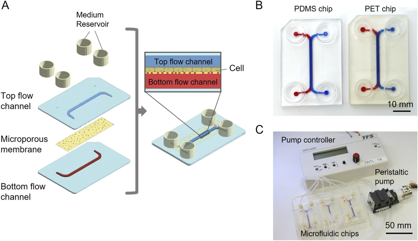
Holding Chip Geometry Constant while Changing Material
Tokai University researchers compared two kidney microphysiological systems with the same channel geometry but different gas permeability. One used polyethylene terephthalate, or PET; the other used polydimethylsiloxane, or PDMS. Conventional culture inserts provided a third comparison. The study investigates how the physical culture setting affects ischemia–reperfusion modeling and the response to CDDO.
All three systems used human RPTEC/TERT1 proximal-tubule epithelial cells. For the injury condition, cultures underwent 24 hours without glucose and without chip perfusion at nominal oxygen settings of 5% or 1%, followed by 24 hours of reoxygenation with glucose-containing medium. Chip perfusion was restored during this second period; inserts remained static.
Finite-element simulations estimated oxygen distribution and consumption in each configuration. The analysis predicted continued oxygen supply through PDMS, whereas PET chips and inserts developed stronger oxygen limitation. These were modeled cell-surface oxygen values, interpreted alongside the biological measurements.
Nominal Oxygen Settings Give Different Tissue Conditions
At the same low-oxygen settings, PDMS cultures retained an intact monolayer and showed no significant corresponding loss of viability. PET chips developed cell detachment and substantial viability loss after the injury/recovery sequence. Under the reported 5% and 1% conditions, viability in PET chips fell by approximately half.
Inserts generally produced milder injury than PET chips. In the insert experiments, low oxygen induced HIF1A, GLUT1 and VEGFA expression. KIM-1 increased under the more severe 1% condition and remained elevated after reoxygenation. Because severe cell loss limited RNA recovery from PET chips, these gene-expression measurements came from inserts and were not presented as direct PET-chip transcript data.
The comparison ties material permeability and medium handling to the biological condition that develops around the cells. Incubator settings alone did not determine whether the intended injury phenotype was produced.
CDDO Responses Depend on the Tested Platform
The pharmacological experiment used CDDO under the 1% oxygen injury condition. CDDO is associated with Nrf2-related cytoprotection in prior work, and the study used its concentration-dependent protective and toxic effects as a test of the culture systems.
In inserts, viability improved with increasing concentration up to approximately 100 nM, where the improvement was significant relative to injured cultures without CDDO. PET-chip measurements showed an improving trend up to 1 μM. At higher concentrations, viability declined, with a significant reduction at 100 μM in inserts and above 10 μM in PET chips.
These observations establish different concentration-response patterns in the two platforms. The authors relate the shift to differences in the starting injury and oxygen environment. They also identify a wider panel of oxygen conditions as necessary for testing whether the protective concentration range changes systematically with oxygen limitation.
Integrating Engineering and Biological Evidence
The drug experiment was performed at one oxygen-deprivation setting, and the study did not directly measure in-chip oxygen with a sensor. Simulations supplied the oxygen estimates; morphology, viability and selected gene measurements supplied experimental support for the resulting biological interpretation.
The cell model is an immortalized proximal-tubule line, and the main outcomes emphasize viability rather than a complete range of renal functions. Broader cellular composition, direct oxygen sensing and more functional endpoints would extend the evaluation. At least three independent experiments supported the principal injury and drug comparisons.
The study illustrates a practical aspect of NAMs assay design: device material, geometry, flow and cell consumption together shape the exposure environment. Connecting that engineering information with the baseline injury and measured drug response helps explain why nominally similar experiments can produce different pharmacological results.