
Low-Flow Culture Preserves Kidney Organoid Structure during Vascularization
A study combines kidney organoids, vascular-supporting cells and collagen scaffolds to compare perfusion conditions. Lower flow preserved nephron-like architecture, while separate tacrolimus experiments measured tissue and vascular injury.
Engineering Support around Kidney Organoids
Researchers at Maastricht University and the University of Basel investigated how vascular-supporting cells, extracellular matrix and perfusion influence human kidney organoids. Their modular co-culture system combines iPSC-derived kidney tissue with stromal vascular fraction cells, collagen sponge scaffolds and optional decellularized kidney matrix. The study treats vascular development and preservation of kidney architecture as related experimental outcomes.
Kidney organoids were generated from a nephron-progenitor-enriched population derived from one human iPSC line. Stromal vascular fraction cells were cultured in collagen scaffolds to form vessel-like networks. Bringing those components together allowed the investigators to compare scaffolds with and without pre-existing vascular structures, rather than attributing every change to perfusion alone.
The Balance between Vascular Networks and Tissue Architecture
Serum supplementation supported endothelial and pericyte-associated structures. In the initial comparison, 1.5% fetal bovine serum was sufficient to sustain vascular networks, while higher supplementation produced stronger vascular-marker responses. The subsequent experiments used the lower level to balance vascular support with preservation of kidney-tissue features.
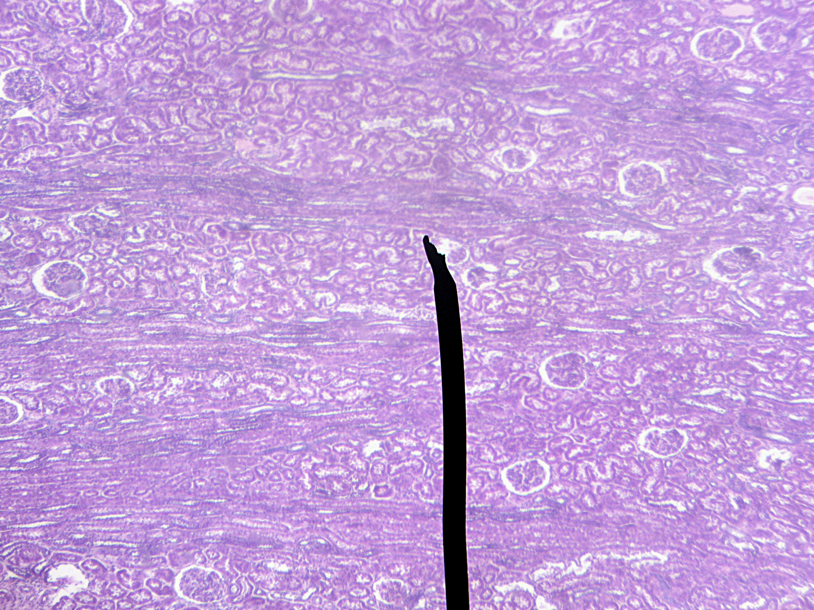
The investigators compared static culture with flow rates of 0.01 and 0.47 mL/min. The lower rate was selected to approximate interstitial tissue flow; the higher rate had previously been optimized for vascularization in the scaffold system. Under the higher-flow condition, organoids lost characteristic morphology and showed disruption of glomerular-like structures. Lower flow better preserved these features while allowing vascular expansion around the tissue.
In the comparison without added decellularized matrix, vessel coverage fraction and total vessel-length density did not significantly increase across the scaffold and perfusion conditions. SVF prevascularization significantly increased vessel endpoint density.
Adding Kidney Extracellular Matrix
Encapsulation in decellularized porcine kidney extracellular matrix preserved glomerular-like domains and supported vascular infiltration. However, matrix-encapsulated organoids were smaller than suspension controls, and some vascular measurements were lower. The matrix condition was then assessed with the scaffold and flow combinations to examine how these components interacted.
Stromal vascular fraction co-culture produced close spatial relationships between endothelial networks and podocyte-associated regions. These images provide information about tissue organization and possible cell interactions. The experiment did not establish a mature perfused glomerular filtration system. The authors describe the culture as a proof of concept for engineering vascularized kidney tissue, with vascular maturation and long-term stability remaining development priorities.
Separating the Tacrolimus Experiments
Tacrolimus was used to test whether the systems could register drug-associated injury. A concentration–response experiment in suspension-grown organoids produced an LDH-based CC50 of 70.87 μM after 24 hours. Increasing exposure was accompanied by tissue breakdown, increased KIM-1 staining and loss of vascular integrity.
Prevascularized SVF scaffolds without kidney organoids showed only about 3% LDH cytotoxicity across 0–100 μM tacrolimus. Organoid-containing co-cultures under low flow showed KIM-1 induction and loss of vascular integrity at 70 μM, near the suspension-organoid CC50. The reported suspension-culture CC50 belongs to the dose–response experiment in that configuration; the vascular co-culture was assessed through its corresponding injury measurements. Keeping the configurations distinct is important when comparing sensitivity or selecting a concentration for a new study.
The pharmacological demonstration used one compound and limited independent replication. Its practical contribution is a set of linked measurements: kidney-tissue structure, vascular organization, stress markers and cytotoxicity. Together with the flow comparisons, those endpoints show how culture design can be evaluated before the system is extended to broader nephrotoxicity testing.