
Autologous TIL–Lung Organoid Coculture Links Killing with Mixed T-Cell States
Patient-matched lung cancer organoids and tumor-infiltrating lymphocytes supported short-term killing assays and single-cell profiling. Cytotoxic changes appeared alongside exhaustion-associated signals, while pathway enrichment remained associative and clinical response prediction was untested.
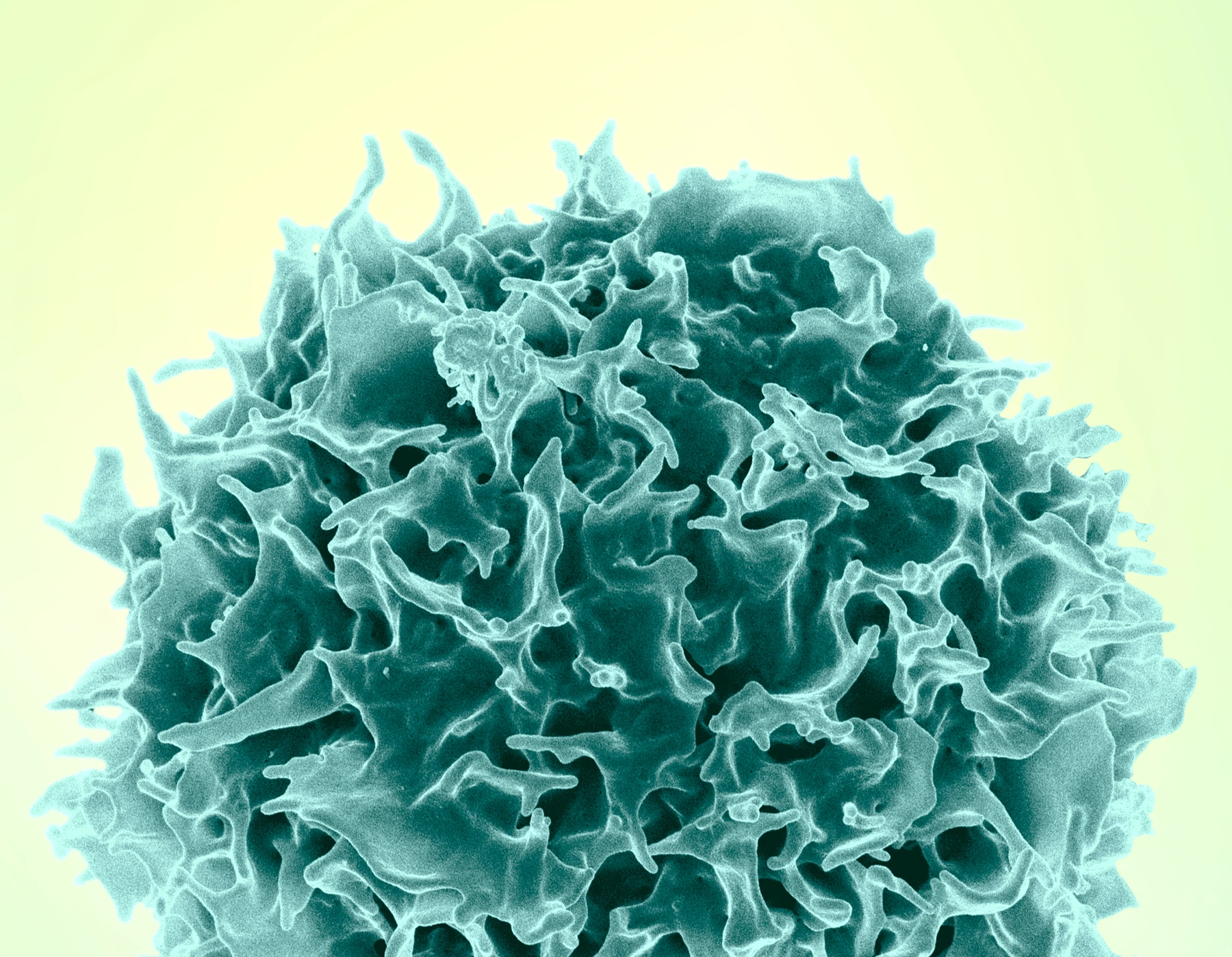
Combining Functional Killing with Immune Profiling
Tumor-infiltrating lymphocytes can contain cancer-reactive T cells, but their activity changes during expansion and contact with a target. A Communications Biology study from Peking University Shenzhen Hospital and collaborating academic centers paired lung cancer organoids with autologous TILs. The researchers measured short-term tumor injury and then profiled the lymphocyte populations associated with coculture.
Organoids formed from twelve patients: eight with lung adenocarcinoma and four with lung squamous cell carcinoma. Different experiments used different subsets. T-cell-receptor repertoire comparisons involved two patients, representative functional killing tests used selected cases, and single-cell profiling used five independent patient samples. These subsets describe separate evidence for culture, function and cell state.
Expansion Narrowed the Receptor Repertoire
Comparing TILs at the start of culture and after 14 days showed fewer unique T-cell-receptor clones and lower Shannon diversity, with enrichment of dominant shared clones. A lung-cancer-associated receptor score also increased. These changes suggest selection during expansion, although a repertoire score does not demonstrate functional tumor recognition by every enriched clone.
Functional experiments paired the expanded lymphocytes with the same patient’s organoids at effector-to-target ratios of 5:1 or 10:1 for 48 hours. Imaging every two to three hours was complemented by caspase-3/7 activity, flow-cytometric target-cell viability, lactate dehydrogenase release and cytokine measurements. The high effector ratios provided a therapeutic testing condition rather than an estimate of the cell balance in a tumor.
Multiple Readouts Supported Selected Autologous Responses
In one representative case, the live CFSE-positive, DAPI-negative target-cell fraction (mean ± standard deviation) was 5.37% ± 3.33% after TIL coculture, compared with 43.10% ± 8.65% in organoids alone. This comparison used three technical replicates and gave P = 0.010. A blood-mononuclear-cell control showed little killing, with a mean live-target fraction of approximately 49.2% in two technical replicates.
LDH measurements also showed greater injury in autologous tumor organoids than in matched normal organoids. In a selected cross-patient comparison, TILs killed their own patient’s tumor more effectively than organoids from two other patients. These tests provide functional evidence of selective recognition in the examined combinations. They do not establish the same magnitude of killing across all twelve patients.
Cytotoxic and Exhaustion-Associated Features Coexisted
Single-cell analysis of five patients resolved 28 broad clusters and 25 T-cell subclusters. Coculture increased GZMB expression, while GZMH did not change. TNF- and IL-17-related pathways were enriched, but no pathway-blocking experiment established that either pathway caused the observed tumor killing.
The relative proportions of CD4 effector-memory and Th2 populations increased, while proliferative CD4-MKI67 and CD8-HAVCR2 populations decreased in the paired analysis. At the same time, exhaustion-associated transcripts including PDCD1, LAG3, HAVCR2, TIGIT and TOX increased. Cell-population proportions and transcript abundance therefore gave a mixed picture rather than a uniform transition toward a more effective or less exhausted state.
The paired single-cell comparisons used five donors and one-tailed tests. Changes in cluster proportions do not trace individual cells through a differentiation sequence. The profiling links cell states to the coculture condition, with their functional contribution requiring further perturbation experiments.
Scope of the 48-Hour Coculture Assay
The organoid–TIL pairing makes target injury and immune state observable within the same patient background. Small experimental subsets, high effector ratios, incomplete tumor microenvironment and a 48-hour functional window limit generalization. No matched clinical TIL-treatment outcomes were used to validate prediction. The assay compares recognition and killing with accompanying T-cell states, including coexisting cytotoxic and exhaustion-associated signals.